. 2013 Mar;20(3):363-70.
doi: 10.1038/nsmb.2500. Epub 2013 Feb 10.

A recurring motif for antibody recognition of the receptor-binding site of influenza hemagglutinin

Affiliations

A recurring motif for antibody recognition of the receptor-binding site of influenza hemagglutinin

Rui Xu et al. Nat Struct Mol Biol. 2013 Mar.

Abstract

Influenza virus hemagglutinin (HA) mediates receptor binding and viral entry during influenza infection. The development of receptor analogs as viral-entry blockers has not been successful, which suggests that sialic acid may not be an ideal scaffold to obtain broad, potent HA inhibitors. Here, we report crystal structures of Fab fragments from three human antibodies that neutralize the 1957 pandemic H2N2 influenza virus in complex with H2 HA. All three antibodies use an aromatic residue to plug a conserved cavity in the HA receptor-binding site. Each antibody interacts with the absolutely conserved HA1 Trp153 at the cavity base through π-π stacking with the signature Phe54 of two VH1-69-encoded antibodies or a tyrosine from HCDR3 in the other antibody. This highly conserved interaction can be used as a starting point to design inhibitors targeting this conserved hydrophobic pocket in influenza viruses.


Conflict of interest statement

Competing financial interests

Vanderbilt University previously submitted a patent covering the diagnostic and therapeutic use of antibodies 2G1, 8M2 and 8F8 prior to the structural work described here.

Figures

Fig. 1
Fig. 1
Crystal structures of H2 HA in complex with Fab 2G1 (a) and Fab 8M2 (b). Three Fabs are associated with each HA trimer. One of the Fabs is colored in blue (heavy chain) and cyan (light chain) and the corresponding HA1 in yellow and HA2 in green. N-linked glycans that are observed in the crystal structure are shown in spheres (carbon in grey, oxygen in red and nitrogen in blue). The other two protomers in the HA trimer and their associated Fabs are colored in light grey.
Fig. 2
Fig. 2
Antibody–antigen binding footprints in the 2G1 and 8M2 complexes. (a) Footprint of 2G1 on the HA. HA residues interacting with the heavy-chain are colored in yellow with the light chain contacts in green. Residues in contact with both heavy and light chains are shown in light green. CDR loops are shown in ribbons. Phe54 on HCDR2 inserts into the receptor-binding pocket and is highlighted in red sticks. (b) Footprint of 8M2 on the HA. HA and CDR loops are colored as in panel A. (c) Footprint of HA on 2G1 combining site. (d) Footprint of HA on 8M2 combining site. Antibody residues in contact with HA are shown in darker colors (heavy chain in blue on a purple background and light chain in cyan on a whitish cyan background).
Fig. 3
Fig. 3
Recognition of the receptor-binding site by the HCDR2 and HCDR3 loops of 2G1 (a) and 8M2 (b). In the crystal structures, antibodies 2G1 and 8M2 approach the HA from completely opposite orientations, with the two HCDR3s interacting on opposite ends of the receptor-binding site, rotated around HCDR2 by approximately 180°. The signature motif of antibodies encoded by germline gene VH1-69 is the hydrophobic tip of HCDR2 loop containing Ile53 and Phe54. In both structures, Phe54 is buried in the conserved hydrophobic pocket formed by Trp153 and neighboring residues of HA1, where sialic acid binds. The rest of the HCDR2 loop and the HCDR3 residues contact different parts of the HA receptor-binding site. Residues key to the interaction are highlighted in sticks, and hydrogen bonds are shown in dashed lines.
Fig. 4
Fig. 4
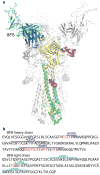
Crystal structure of 8F8 with H2 HA. (a) Overall structure of the antibody–antigen complex. Three Fabs bind to the HA trimer. Fab 8F8 (heavy chain in blue, light chain in cyan) targets the receptor-binding site of HA (HA1 in yellow and HA2 in green). N-linked glycans that are observed in the crystal structure are shown in spheres (carbon in grey, oxygen in red and nitrogen in blue). The other two protomers in the HA trimer and their associated Fabs are colored in light grey. (b) Amino-acid sequences of 8F8 fragment. Residues that are in contact with HA in the complex structure are highlighted in red. 8F8 binding to H2 HA is dominated by the heavy chain.
Fig. 5
Fig. 5
Antibody–antigen recognition by 8F8. (a) Footprint of 8F8 on the HA. HA residues interacting with the heavy chain are colored in yellow and with the light chain in green. Residues in contact with both heavy and light chains are shown in light green (residue 189). CDR loops are shown in ribbons. Tyr100 (shown as red sticks) on the HCDR3 inserts into a hydrophobic pocket in the receptor-binding site. (b) Footprint of HA on 8F8 combining site. Antibody residues in contact with HA are shown in darker colors (heavy chain in blue on a purple background and light chain in cyan on a whitish cyan background). (c) At the antibody–antigen interface in the crystal structure, the 8F8 HCDR3 loop dominates the interactions with HA. Residues key to the antibody association are highlighted in sticks and hydrogen bonds are shown in dashed lines. Tyr100forms π-π interactions with HA Trp153. (d) Comparison between 8F8 and CH65, an antibody targeting the receptor-binding site of seasonal H1 HA. In the structure of CH65 (ref. ), Val100B, instead of Tyr100 in 8F8, occupies the hydrophobic pocket in the receptor-binding site.
Fig. 6
Fig. 6
Escape mutations on H2 HA for binding to antibodies 8F8, 8M2 and 2G1 and their effects on binding of glycan receptors. (a) Escape mutants were identified in vitro: R137Q or T193K for 8F8, G135D for 8M2 and K156E for 2G1. (b) Glycan binding analysis of recombinant wild-type (WT) H2 HA on glycan microarray. WT H2 HA shows specific binding toward certain α2–6-linked sialylated glycans (red bars; 36 to 56) and glycans of α2–6 and α2–3 mixed linkages (cyan; 57 and 58), but not to α2–3-linked glycans (blue; 3 to 35) or neutral glycans (black; 1 and 2). The list of glycans on the array is provided in Supplementary Table 1. (c) The single mutation R137Q abolishes glycan binding (or weakens glycan binding to below the detection threshold) for H2 HA, compared with the WT HA tested under the same conditions. All error bars in the figure are indicative of standard deviation from quadruplicates. Similar results for mutations T193K, G135D or K156E are included in Supplementary Fig. 2.
Fig. 7
Fig. 7
Targeting the receptor-binding site. (a) Conservation of a key interaction for three H2 antibodies targeting the receptor-binding site of HA. Aromatic residues at the tip of CDR loops (Tyr100 of 8F8 in yellow, Phe54 of 8M2 in green, Phe54 of 2G1 in cyan) insert into the highly conserved hydrophobic pocket of HA in the same pocket that sialic acid of the glycan receptor binds (HA is shown in electrostatic surface representation (positive charges in blue, negative charges in red and neutral in white). (b) The location for binding of sialic acid in the receptor-binding site was modeled based on the crystal structure of H2 HA in complex with human receptor analog LS-Tetrasaccharide c (LSTc) (PDB code: 2WRE ).

Comment in

  • Host versus flu: antibodies win a round?
    Brooke CB, Yewdell JW. Brooke CB, et al. Nat Struct Mol Biol. 2013 Mar;20(3):245-6. doi: 10.1038/nsmb.2524. Nat Struct Mol Biol. 2013. PMID: 23463305 Free PMC article.

References

    1. Gilbert SC. Advances in the development of universal influenza vaccines. Influenza Other Respi Viruses. 2012 - PMC - PubMed
    1. Ekiert DC, Wilson IA. Broadly neutralizing antibodies against influenza virus and prospects for universal therapies. Curr Opin Virol. 2012;2:134–41. - PMC - PubMed
    1. Du J, Cross TA, Zhou HX. Recent progress in structure-based anti-influenza drug design. Drug Discov Today. 2012;17:1111–20. - PMC - PubMed
    1. Skehel J, Wiley D. Receptor binding and membrane fusion in virus entry: the influenza hemagglutinin. Annu Rev Biochem. 2000;69:531–69. - PubMed
    1. Paulson JC. The Receptors. Vol. 2. Academic Press; Orlando, FL: 1985. Interactions of animal viruses with cell surface receptors; pp. 131–219.

Publication types

MeSH terms