. 2013 Mar;20(3):347-54.
doi: 10.1038/nsmb.2501. Epub 2013 Feb 10.

Human RECQ1 promotes restart of replication forks reversed by DNA topoisomerase I inhibition

Affiliations

Human RECQ1 promotes restart of replication forks reversed by DNA topoisomerase I inhibition

Matteo Berti et al. Nat Struct Mol Biol. 2013 Mar.

Abstract

Topoisomerase I (TOP1) inhibitors are an important class of anticancer drugs. The cytotoxicity of TOP1 inhibitors can be modulated by replication fork reversal through a process that requires poly(ADP-ribose) polymerase (PARP) activity. Whether regressed forks can efficiently restart and what factors are required to restart fork progression after fork reversal are still unknown. We have combined biochemical and EM approaches with single-molecule DNA fiber analysis to identify a key role for human RECQ1 helicase in replication fork restart after TOP1 inhibition that is not shared by other human RecQ proteins. We show that the poly(ADP-ribosyl)ation activity of PARP1 stabilizes forks in the regressed state by limiting their restart by RECQ1. These studies provide new mechanistic insights into the roles of RECQ1 and PARP in DNA replication and offer molecular perspectives to potentiate chemotherapeutic regimens based on TOP1 inhibition.


Figures
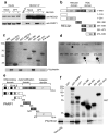
Figure 1
Figure 1. Analysis of the RECQ1-PARP1 interaction
(a) IPs from U-2 OS cells using the anti-RECQ1 antibody ± PARP inhibitor (50 μM NU1025) and ± DNA damage (100 nM CPT for 2 hrs). (b) Schematic representation of the domain structure of RECQ1 and the GST-tagged RECQ1 fragments (D1 and D2 are the RecA-like domains). (c) Pull-down assays with GST-tagged RECQ1 fragments. Top: Coomassie stained gel of GST-RECQ1 fragments. Bottom: autoradiography of in vitro GST pull-down assay using 35S-labeled PARP1 protein. (d) Analysis of PAR binding in vitro. RECQ1 fragments (2 pmol) were dot-blotted onto a nitrocellulose membrane and incubated with 32P-labeled PAR. (e) Schematic representation of the domain structure of PARP1 and the GST-tagged PARP1 fragments (A, DNA binding domain; B, nuclear localization signal; D, BRCT–automodification domain; E, contains a WGR motif; F, catalytic domain. A third zinc-finger motif has been recently identified in domain C , in addition to the previously identified FI and FII zinc-finger motifs. NLS is a nuclear localization sequence. (f) Pull-down assays with GST-tagged PARP1 fragments. Bound proteins were revealed by autoradiography (bottom panel). Purified GST or GST–PARP1 proteins were detected with an anti-GST antibody (top panel). Input: 20% of the amount used in binding reactions.
Figure 2
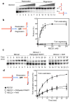
Figure 2. The in vitro fork restoration activity of RECQ1 is inhibited by PARylatedPARP1 and PAR
(a) Lanes 1–7: fork restoration assays performed using increasing RECQ1 concentrations (0, 15, 25, 35, 50, 100, and 200 nM) and a fixed concentration of the chicken foot substrate (2 nM). Lanes 8–14: fork regression assays using increasing RECQ1 concentrations (0, 15, 25, 35, 50, 100, and 200 nM) and a fixed concentration of the replication fork structure (2 nM). The hatched regions indicate heterologous sequences that are included in the vertical arms to prevent complete strand separation. In addition, we inserted two mismatches and a single isocytosine modification to prevent spontaneous fork regression and restoration (see Supplementary Fig. 3 for more details). All the reactions were stopped after 20 min. (b) Left: reaction scheme. Right: Plot of the fork restoration and regression activities as a function of protein concentration. (c) Lanes 1–7: kinetic experiments performed using 40 nM RECQ1 and the chicken foot substrate (2 nM). Lanes 8–14: kinetic experiments performed in the presence of PARylatedPARP1 (40 nM). Lanes 15–21: kinetic experiments performed in the presence of PAR (100 nM). (d) Left: reaction scheme. Right: Plots of the fork restoration assays performed in the presence and absence of PARylatedPARP1 or PAR. The data points in a, b, c, and d represent the mean of three independent experiments. Error bars indicate standard error of the mean (s.e.m).
Figure 3
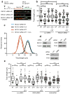
Figure 3. Restoration of normal replication fork progression after TOP1 and PARP inhibition is impaired in RECQ1-depleted—but not in WRN- or BLM-depleted—U-2 OS cells
(a) Schematic of single DNA fiber replication track analysis. U-2 OS cells were transfected with siRNA against luciferase (Luc) or RECQ1 before CldU or IdU labeling, as indicated. Red and green denote CldU- and IdU-containing tracts, respectively. 50 nM CPT has been added concomitantly with the second label. Representative DNA fiber tracts from Microfluidic-assisted replication tract analysis of RECQ1 depleted U-2 OS cells upon TOP1 and/or PARP inhibition are shown below. White scale bar represents 12.5 μm long. (b) Statistical analysis of IdU tract length measurements from Luc- or RECQ1-depleted cells. Relative length of IdU tracts (green) synthesized after mock (NT) or CPT treatment (50nM). At least 175 tracts were scored for each dataset. 10 μM Olaparip (OLA) was optionally added 2 hours before CldU labeling and maintained during labeling. Whiskers indicate the 10th and 90th percentiles. Statistical test according to Mann-Whitney, results are ns not significant, **p 0.006, **** p<0.0001. (c) The smoothed histogram shows the green (IdU) tract length distribution after TOP1 and/or PARP inhibition in RECQ1-downregulated cells. (d) Western blot for RECQ1, BLM, and WRN downregulation. (e) Statistical analysis of IdU tract length measurements from Luc-, WRN-, or BLM-depleted cells performed as described in b.
Figure 4
Figure 4. Genetic complementation of RECQ1-depleted cells with wild-type RECQ1, but not with the ATP-deficient K119R mutant, rescues the fork progression phenotype observed in RECQ1-depleted U-2 OS cells
(a) Experimental scheme for the genetic knockdown-rescue experiments. U-2 OS cells were transduced with lentivirus to luciferase (Luc shRNA) or RECQ1 (RECQ1 shRNA). RECQ1-depleted cells were genetically complemented by nucleofection of RNAi resistant vectors, for the expression of wild-type (WT-shR) and ATPase-deficient (K119R-shR) shRNA resistant forms of RECQ1 before CldU labeling, as indicated. (b) Western blot analysis of RECQ1-depleted cells complemented with the shRNA resistant wild type RECQ1 or K119R mutant (both proteins are Flag-tagged). Tubulin was detected as a loading control. (c) Statistical analysis of IdU tract length measurements from a. Relative length of IdU tracts (green) synthesized after CPT treatment (50 nM). At least 175 tracts were scored for each dataset. 10 μM Olaparip was added 2 hours before CldU labeling and maintained during labeling. Whiskers indicate the 10th and 90th percentiles. Statistical test according to Mann-Whitney, results are ns not significant, **** p<0.0001. (d) The smoothened histogram shows green (IdU) tract length after Top1 and PARP inhibition in RECQ1-downregulated cells and RECQ1-downregulated cells genetically complemented with wild-type RECQ1 or the ATPase deficient K119R mutant.
Figure 5
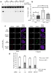
Figure 5. PARP inactivation leads to DSB formation at low CPT doses in the presence, but not in the absence of RECQ1
(a) PFGE of U-2 OS cells transfected with siRNA against Luc and RECQ1, and treated with either CPT (100 nM) and/or Olaparib (10 μM). (b) DSB signals were quantified by ImageJ and normalized to unsaturated signals of DNA retained in the wells. The values obtained from the different treatments were then normalized against their respective untreated controls to obtain the fold change in DSBs upon treatment with CPT. The graph integrates results from three independent experiments. Statistical analysis was done using paired t test (* represents significance level where p value <0.05, ns represents non-significance between the samples) (c) Immunofluorescence experiments of U-2 OS cells were treated with 100 nM CPT, as indicated, ± 10 μM Olaparib, and co-stained for γH2AX and 53BP1. A representative image is shown in each condition. (d) The plot shows the average number of γH2AX foci per cell and the average fraction (± s.e.m.) of γH2AX foci colocalizing with 53BP1.
Figure 6
Figure 6. Reversed forks accumulate and are unable to restart in RECQ1-depleted cells after CPT treatment even if PARP is inhibited
(a) Representative electron micrograph of a reversed fork observed on genomic DNA from CPT (25 nM) + Olaparib (10 μM) treated U-2 OS cells transfected with siRECQ1. The white arrow points to the four-way junction at the replication fork. D = Daughter strand, P = Parental strand, R = Reversed arm. (b) Frequency of fork reversal in U-2 OS cells transfected either with siLuc or siRECQ1 and treated with CPT ± Olaparib. Restart experiments were performed measuring the frequency of fork reversal 3 hours after CPT removal. In brackets (down), the number of analyzed molecules, and (up) the % of reversed forks.
Figure 7
Figure 7. Schematic model of the combined roles of PARP1 and RECQ1 in response to TOP1 inhibition
(a, b) PARP poly(ADPribosyl)ation activity is not required to form reversed forks, but it promotes the accumulation of regressed forks by inhibiting RECQ1 fork restoration activity, thus preventing premature restart of the regressed forks. (c) Inhibition of PARP activity leads to replication run-off and increased DSBs formation upon TOP1 inhibition since RECQ1 can restart reversed forks untimely. (d) PARP activity is no longer required in RECQ1-depleted cells were regressed forks accumulate because the cells lack the enzyme (RECQ1) necessary to promote fork restart. HR might be required to promote fork restart in the absence of RECQ1 and PARP activity.

References

    1. Koster DA, Palle K, Bot ES, Bjornsti MA, Dekker NH. Antitumour drugs impede DNA uncoiling by topoisomerase I. Nature. 2007;448:213–7. - PubMed
    1. Pommier Y, Leo E, Zhang H, Marchand C. DNA topoisomerases and their poisoning by anticancer and antibacterial drugs. Chemistry & biology. 2010;17:421–33. - PMC - PubMed
    1. Pommier Y. Topoisomerase I inhibitors: camptothecins and beyond. Nature reviews Cancer. 2006;6:789–802. - PubMed
    1. Rodriguez-Galindo C, et al. Clinical use of topoisomerase I inhibitors in anticancer treatment. Medical and pediatric oncology. 2000;35:385–402. - PubMed
    1. Pommier Y, et al. Repair of and checkpoint response to topoisomerase I-mediated DNA damage. Mutation research. 2003;532:173–203. - PubMed

Publication types

MeSH terms