Review
. 2013 Apr 15;304(8):H1060-76.
doi: 10.1152/ajpheart.00646.2012. Epub 2013 Feb 8.

Ketone body metabolism and cardiovascular disease

Affiliations
Review

Ketone body metabolism and cardiovascular disease

David G Cotter et al. Am J Physiol Heart Circ Physiol. .

Abstract

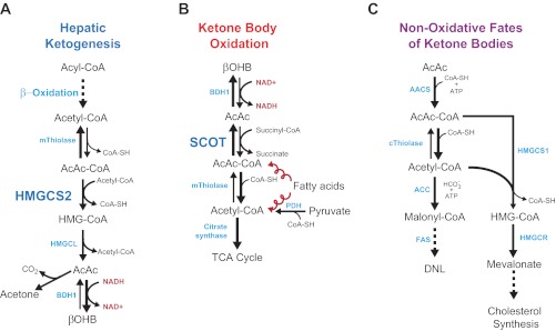
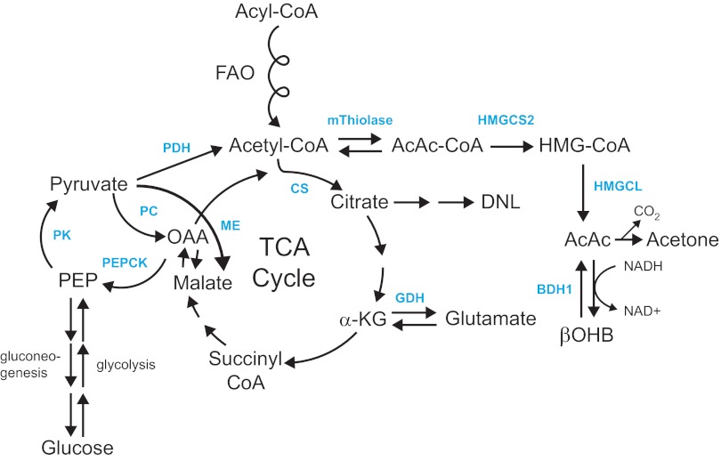
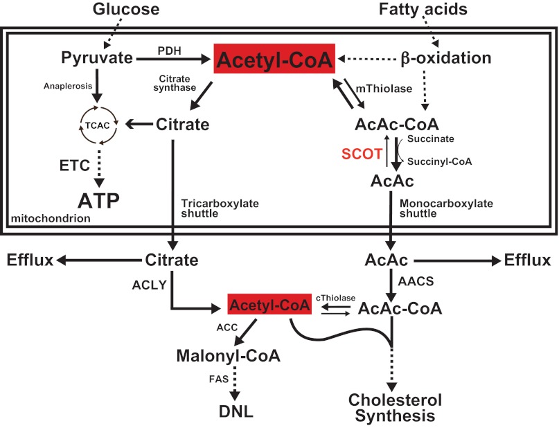
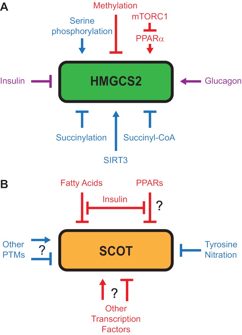
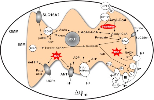
Ketone bodies are metabolized through evolutionarily conserved pathways that support bioenergetic homeostasis, particularly in brain, heart, and skeletal muscle when carbohydrates are in short supply. The metabolism of ketone bodies interfaces with the tricarboxylic acid cycle, β-oxidation of fatty acids, de novo lipogenesis, sterol biosynthesis, glucose metabolism, the mitochondrial electron transport chain, hormonal signaling, intracellular signal transduction pathways, and the microbiome. Here we review the mechanisms through which ketone bodies are metabolized and how their signals are transmitted. We focus on the roles this metabolic pathway may play in cardiovascular disease states, the bioenergetic benefits of myocardial ketone body oxidation, and prospective interactions among ketone body metabolism, obesity, metabolic syndrome, and atherosclerosis. Ketone body metabolism is noninvasively quantifiable in humans and is responsive to nutritional interventions. Therefore, further investigation of this pathway in disease models and in humans may ultimately yield tailored diagnostic strategies and therapies for specific pathological states.


Figures

Fig. 1.
Fig. 1.
Ketone body metabolism pathways. A: ketogenesis within hepatic mitochondria is the primary source of circulating ketone bodies. B: primary metabolic fate of ketone bodies is terminal oxidation within mitochondria of extrahepatic tissues via CoA transferase [succinyl-CoA:3-oxoacid-CoA transferase (SCOT)]. Substrate competition with pyruvate-derived and fatty acyl-CoA-derived (the corkscrew arrows represent the activities of the β-oxidation spiral) acetyl-CoA are shown. C: cytoplasmic de novo lipogenesis (DNL) and cholesterol synthesis are nonoxidative metabolic fates of ketone bodies. For simplicity of C, only acetoacetate (AcAc) is depicted, although β-hydroxybutyrate (βOHB) is also a substrate for lipogenesis after it has been oxidized to AcAc via mitochondrial βOHB dehydrogenase (BDH1). AACS, acetoacetyl-CoA synthetase; ACC, acetyl-CoA carboxylase; AcAc-CoA, acetoacetyl-CoA; ATP, adenosine triphosphate; CoA-SH, free coenzyme A; FAS, fatty acid synthase; HMG-CoA, 3-hydroxymethylglutaryl-CoA; HMGCL, HMG-CoA lyase; HMGCS1, cytoplasmic HMG-CoA synthase; HMGCS2, mitochondrial HMG-CoA synthase; HMGCR, HMG-CoA reductase; NAD+(H), nicotinamide adenine dinucleotide oxidized (reduced); PDH, pyruvate dehydrogenase; TCA, tricarboxylic acid; mThiolase, mitochondrial thiolase; cThiolase, cytoplasmic thiolase. Thiolase activity is encoded by at least 6 genes: ACAA1, ACAA2 (encoding an enzyme known as T1 or CT), ACAT1 (encoding T2), ACAT2, HADHA, and HADHB.
Fig. 2.
Fig. 2.
Hepatic integration of ketogenesis. Integration of hepatic ketogenesis with hepatic TCA cycle, DNL, and pyruvate cycling/glucose metabolism. FAO, β-oxidation of fatty acids; GDH, glutamate dehydrogenase; OAA, oxaloacetate; ME, malic enzyme; PEPCK, phosphenolpyruvate carboxykinase; PC, pyruvate carboxylase; PK, pyruvate kinase; PEP, phosphenol pyruvate; CS, citrate synthase; α-KG, α-ketoglutarate.
Fig. 3.
Fig. 3.
Ketogenic flux through CoA transferase. Because CoA transferase catalyzes an equilibrium reaction, select spatiotemporal metabolic conditions may favor channeling of mitochondrial acetyl-CoA to AcAc, generating a monocarboxylate shuttle that complements the citrate-dependent tricarboxylate shuttle and permits mitochondrial efflux of β-oxidation-derived acetyl-CoA independent of the TCA cycle (TCAC). ACLY, ATP-citrate lyase; ETC, electron transport chain.
Fig. 4.
Fig. 4.
Regulatory mechanisms for HMGCS2 and CoA transferase (SCOT). HMGCS2 (A) and SCOT (B) both undergo transcriptional (red) and post-translational modes of regulation (blue). Some regulatory factors exhibit both classes of effects (violet). mTORC1, mammalian target of rapamycin complex 1; PPARα, peroxisome proliferator-activated receptor-α; PTM; post-translational modification; SIRT3, sirtuin (silent mating type information regulation 2 homolog) 3.
Fig. 5.
Fig. 5.
Diverse roles of CoA transferase in mitochondrial function. βOHB and AcAc cross the plasma membrane via solute ligand carrier protein (SLC) 16A (SLC16A) family members and may employ these or other transporters to enter the mitochondrial matrix. Within the mitochondria, d-βOHB is oxidized to AcAc by the inner-membrane bound and phosphatidylcholine-dependent BDH1. CoA transferase (SCOT) catalyzes a near equilibrium reaction through which CoA is exchanged between succinate and AcAc. Oxidation of ketone bodies occurs by mass action and may diminish reactive oxygen species (ROS) formation, compared with oxidation of fatty acids. See text for details. ADP, adenosine diphosphate; ANT, adenine nucleotide transporter; CPT, carnitine palmitoyltransferase; Cyt C, cytochrome c; e, electron; H+, hydrogen ion; I, II, II, IV, complexes I-IV of the electron transport chain, respectively; IMM, inner mitochondrial membrane; OMM, outer mitochondrial membrane; Q, ubiquinone; Δψm, electrochemical potential across the inner mitochondrial membrane; Tr, translocase; UCPs, uncoupling proteins; FAD, flavin adenine dinucleotide.

References

    1. Abel ED, Doenst T. Mitochondrial adaptations to physiological versus pathological cardiac hypertrophy. Cardiovasc Res 90: 230–242, 2011 - PMC - PubMed
    1. Aguilo F, Camarero N, Relat J, Marrero PF, Haro D. Transcriptional regulation of the human acetoacetyl-CoA synthetase gene by PPARgamma. Biochem J 427: 255–264, 2010 - PubMed
    1. Ahmed K, Tunaru S, Offermanns S. GPR109A, GPR109B and GPR81, a family of hydroxy-carboxylic acid receptors. Trends Pharmacol Sci 30: 557–562, 2009 - PubMed
    1. Al-Zaid NS, Dashti HM, Mathew TC, Juggi JS. Low carbohydrate ketogenic diet enhances cardiac tolerance to global ischaemia. Acta Cardiol 62: 381–389, 2007 - PubMed
    1. Aledo R, Zschocke J, Pie J, Mir C, Fiesel S, Mayatepek E, Hoffmann GF, Casals N, Hegardt FG. Genetic basis of mitochondrial HMG-CoA synthase deficiency. Hum Genet 109: 19–23, 2001 - PubMed

Publication types