

Transrectal ultrasound in patients with hematospermia
- PMID: 23396895
- PMCID: PMC3553209
- DOI: 10.1016/j.jus.2009.09.005
Transrectal ultrasound in patients with hematospermia
Abstract
Introduction: To illustrate the lesions detected with transrectal ultrasound (TRUS) in patients with hematospermia.
Material and methods: This study included 74 male patients (25-73 years old) affected by hematospermia. Clinical history was obtained and all patients underwent rectal examination as well as TRUS examination in both axial and coronal planes to evaluate the prostate, ejaculatory ducts and seminal vesicles. Biopsy was performed in 10 patients.
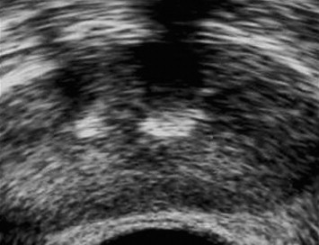
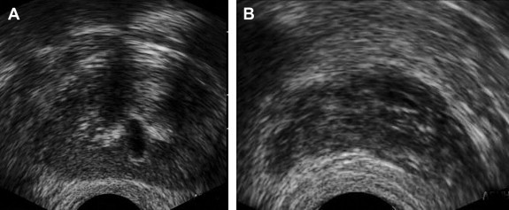
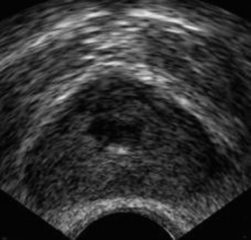
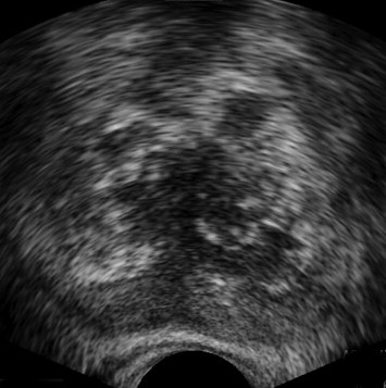
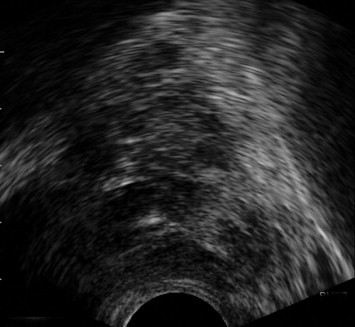
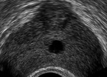
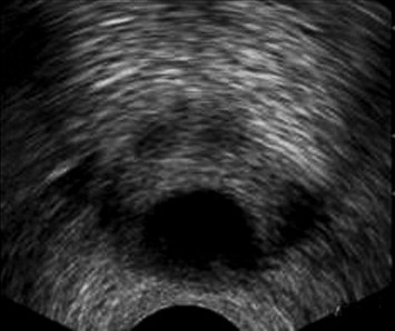
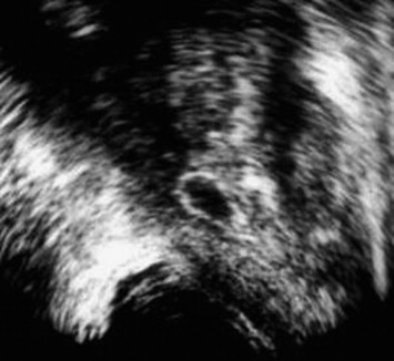
Results: Abnormalities were detected in 59 patients. Calculi (n = 20) were seen within the prostate, seminal vesicles and along the course of the ejaculatory ducts. Chronic prostatitis (n = 14) appeared as hyperechoic and hypoechoic areas within the prostate with capsule thickening suggesting seminal vesiculitis (n = 8). Granulomatous prostatitis (n = 3) appeared as hyperechoic and calcified areas scattered within the prostate and the seminal vesicles. Hypoechoic focal lesions and heterogeneous texture were seen in prostate cancer (n = 5). Utricular cysts (n = 3) appeared as small midline lesions, and Mullerian duct cysts (n = 8) appeared as larger midline cysts protruding above the prostate. Ejaculatory duct cysts (n = 4) appeared as thick walled cystic lesions along the course of the ejaculatory duct. Seminal vesicle cysts were detected in 2 patients.
Conclusion: Our conclusion is that TRUS is a safe, non-invasive technique which can be used to detect lesions of the prostate, seminal vesicles and the ejaculatory ducts in patients with hematospermia.
Sommario INTRODUZIONE: Illustrare le lesioni identificate con ecografia prostatica transrettale in pazienti con ematospermia. MATERIALI E METODI: Il presente studio include 74 pazienti di sesso maschile (25–73 anni) con ematospermia. Tutti i pazienti successivamente sono stati sottoposti a esame ecografico prostatico transrettale sui piani assiale e coronale della prostata, dei dotti eiaculatori e delle vescichette seminali dopo aver effettuato l'anamnesi e l'esplorazione rettale. La biopsia è stata effettuata in 10 pazienti. RISULTATI: In 59 pazienti sono stati identificati reperti patologici. Calcoli (n = 20) sono stati identificati entro il parenchima ghiandolare prostatico, nelle vescichette seminali e lungo il decorso dei dotti eiaculatori. La prostatite cronica (n = 14) si presentava con un'immagine iperecogena con aree ipoecogene del parenchima ghiandolare con ispessimento della capsula che si associava con vesciculite seminale (n = 8). La prostatite granulomatosa (n = 3) appariva iperecogena con alcune regioni calcifiche distribuite nella prostata e nelle vescichette seminali. Nei casi di tumore della prostata sono stati identificate lesioni focali ipoecogene e ecostruttura eterogenea (n = 5). La cisti utriculare (n = 3) era una lesione della linea mediana e le cisti dei Dotti Mulleriani (n = 8) apparivano come una cisti della linea centrale più grande che protrudeva sopra la prostata. Le cisti dei dotti eiaculatori (n = 4) apparivano come lesioni con pareti ispessite lungo il decorso del dotto eiaculatorio e le cisti vescicali sono state identificate in 2 pazienti. CONCLUSIONI: Concludiamo che l'ecografia transrettale è una tecnica non invasiva che può essere usata per identificare lesioni della prostata, delle vescichette seminali e dei dotti eiaculatori in pazienti con ematospermia.
Keywords: Hematospermia; Prostatitis; Ultrasound.
Figures
References
-
- Ahmad I., Krishna N. Hemospermia. J Urol. 2007;177:1613–1618. - PubMed
-
- Munkelwitz R., Krasnokutsky S., Lie J., Shah S.M., Bayshtok J., Khan S.A. Current perspectives on hematospermia: a review. J Androl. 1997;18:6–14. - PubMed
-
- Torigian D.A., Ramchandani P. Hematospermia: imaging findings. Abdom Imaging. 2007;32:29–49. - PubMed
-
- Maeda H., Toyooka N., Kinukawa T., Hattori R., Furukawa T. Magnetic resonance images of haematospermia. Urology. 1993;41:499–504. - PubMed
LinkOut - more resources
Full Text Sources