

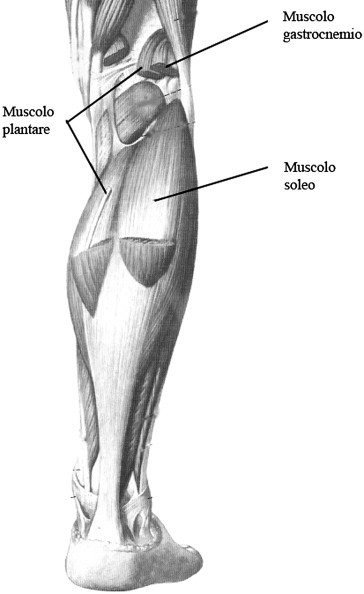
US evaluation and diagnosis of rupture of the medial head of the gastrocnemius (tennis leg)
- PMID: 23396898
- PMCID: PMC3553076
- DOI: 10.1016/j.jus.2007.09.007
US evaluation and diagnosis of rupture of the medial head of the gastrocnemius (tennis leg)
Abstract
Purpose: The aim of this study is to demonstrate the diagnostic accuracy of ultrasonography (US) in the diagnosis of rupture of the medial head of the gastrocnemius muscle, also called "tennis leg" (TL).
Materials and methods: Thirty-five consecutive patients with acute traumatic injury of the calf underwent US examination. There were 25 men and 10 women; mean age 47.5 years (range 35-60 years). All examinations were performed using a 5-12 MHz broadband electronic linear array probe.
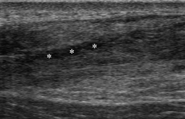
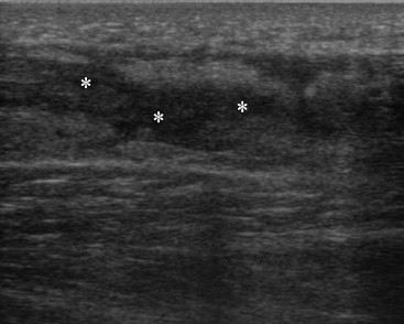
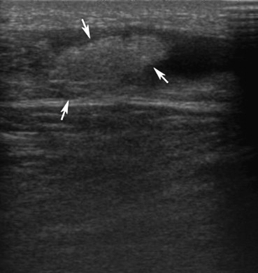
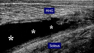
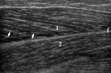
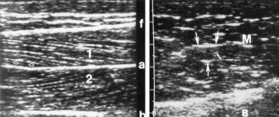
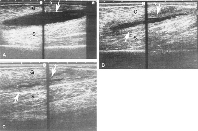
Results: Thirty-three out of 35 patients had TL; 24 cases of partial rupture and nine cases of complete rupture were diagnosed. In the remaining two cases, both with symptoms suggesting TL, one patient had a tear of the proximal musculotendinous junction and one had a ruptured Baker's cyst. Fluid collections caused by the muscular rupture were visible as hypoechoic areas; in 80% of cases associated by a hyperechoic oval area due to hematoma and local inflammation. The degree of fluid collection in the patients with complete rupture (6-16 mm; mean: 9.7 mm) was significantly greater than the one seen in patients with partial rupture (4-8 mm; mean: 6.8 mm).
Conclusions: US is the imaging modality of choice in clinical suspicion of TL, both in the initial workup of the patient and in the follow-up. US is easy to perform and is particularly useful to distinguish TL from other pathologies, especially ruptured Baker's cyst and deep vein thrombosis, which require a different therapeutic management.
Sommario SCOPO: L'obiettivo di tale studio è dimostrare l'accuratezza diagnostica dell'ecografia nella diagnosi della rottura del ventre mediale del muscolo gastrocnemio (Tennis Leg). MATERIALI E METODI: Sono stati valutati con esame ecografico 35 pazienti (25 uomini e 10 donne, con un range di età tra i 35 e i 60 anni ed età media di 47,5 anni) con danno acuto traumatico del polpaccio. Gli esami sono stati effettuati usando una sonda elettronica lineare a banda larga da 5–12 MHz. RISULTATI: Trentatre dei 35 pazienti presentavano una rottura del ventre mediale del muscolo gastrocnemio (TL): 24 rotture parziali e 9 rotture complete. Nei rimanenti due casi, entrambi con sintomi clinici suggestivi di Tennis Leg, sono stati diagnosticati strappo della giunzione prossimale muscolo-tendinea e rottura di una cisti di Baker. La raccolta fluida, conseguenza della rottura muscolare, si presentava come una zona ipoecogena, con area iperecogena ovalare nel contesto, riferibile a ematoma e alla flogosi locale. L'entità della raccolta fluida nei pazienti con rottura completa (6–16 mm; media: 9.7) era significativamente superiore rispetto ai pazienti con rottura parziale (4–8 mm; media: 6.8). CONCLUSIONI: L'ecografia è l'indagine di prima scelta nel sospetto clinico di rottura del ventre mediale del muscolo gastrocnemio, non solo per la rapidità di esecuzione e per il follow-up, ma soprattutto per la possibilità di fare diagnosi differenziale con altre patologie, prime tra tutte la rottura di cisti di Baker e le trombosi venose, che richiedono approccio terapeutico diverso.
Keywords: Gastrocnemius muscle; Tennis leg; Ultrasonography.
Figures
References
-
- Bianchi S., Martinoli C., Fikry Abdelwahab I., Derchi L.E., Damiani S. Sonographic evaluation of tears of the gastrocnemius medial head (“tennis leg”) J Ultrasound Med. 1998;17:157–162. - PubMed
-
- Jarolem K.L., Wolinsky P.R., Savenor A., Ben-Yishay A. Tennis leg leading to acute compartment syndrome. Orthopedics. 1994;17:721–723. - PubMed
-
- Slawski D.P. Deep venous thrombosis complicating rupture of the medial head of the gastrocnemius muscle. J Orthop Trauma. 1994;8:263–264. - PubMed
-
- Gaulrapp H. “Tennis leg”: ultrasound differential diagnosis and follow up. Sportverletz Sportschaden. 1999;13(2):53–58. - PubMed
-
- McClure J.C. Gastrocnemius musculotendinous rupture: a condition confused with thrombophlebitis. South Med J. 1984;77:1143–1145. - PubMed
LinkOut - more resources
Full Text Sources