. 2013 Mar 29;288(13):9438-46.
doi: 10.1074/jbc.M112.448282. Epub 2013 Feb 8.

Atypical protein kinase Cι is required for Wnt3a-dependent neurite outgrowth and binds to phosphorylated dishevelled 2

Affiliations

Atypical protein kinase Cι is required for Wnt3a-dependent neurite outgrowth and binds to phosphorylated dishevelled 2

Yoshimi Endo Greer et al. J Biol Chem. .

Abstract

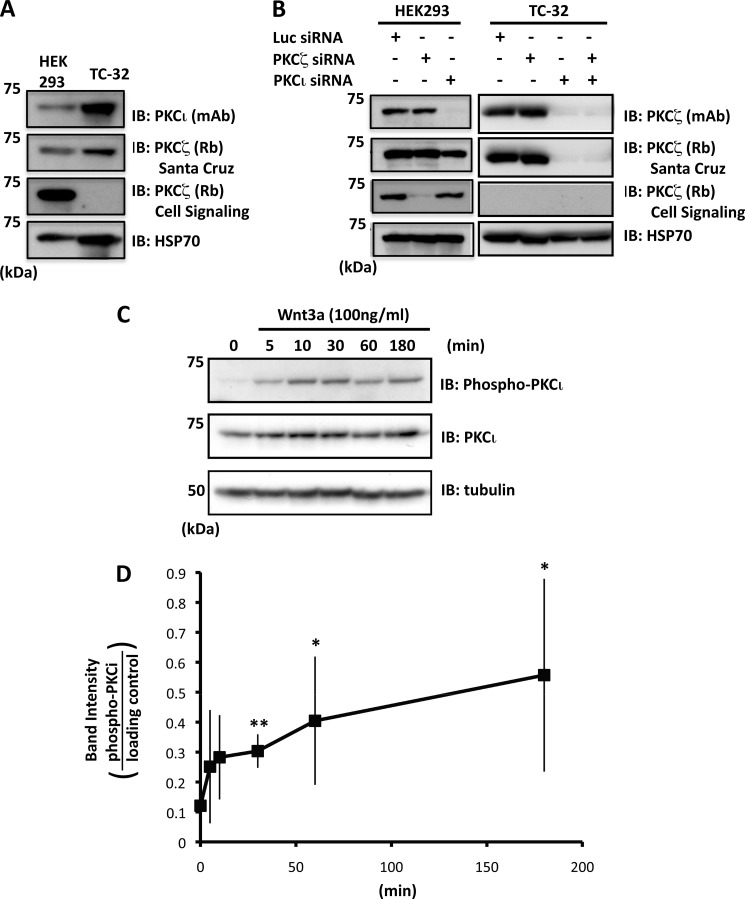
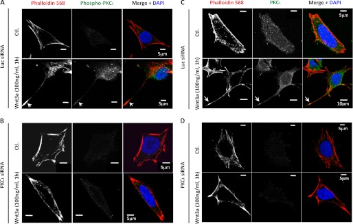
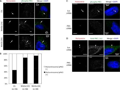
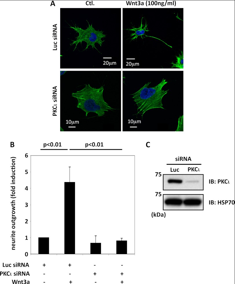
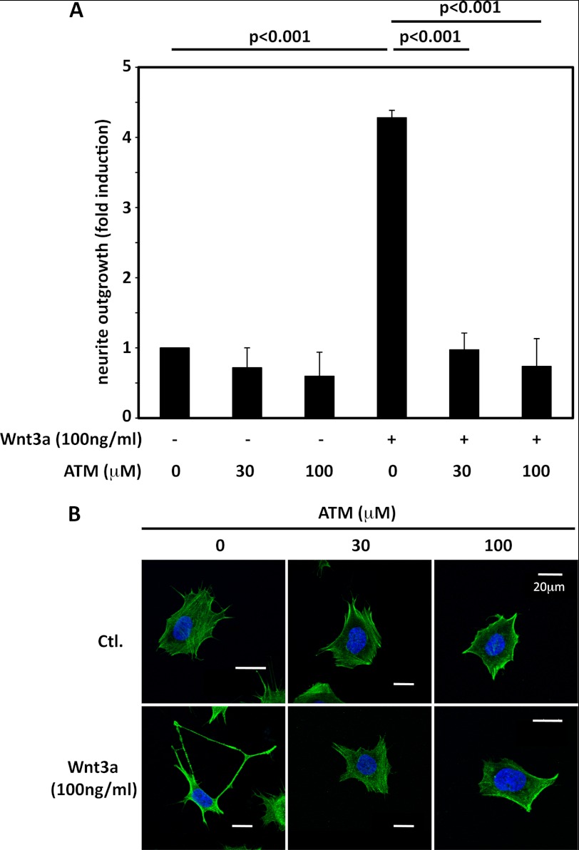
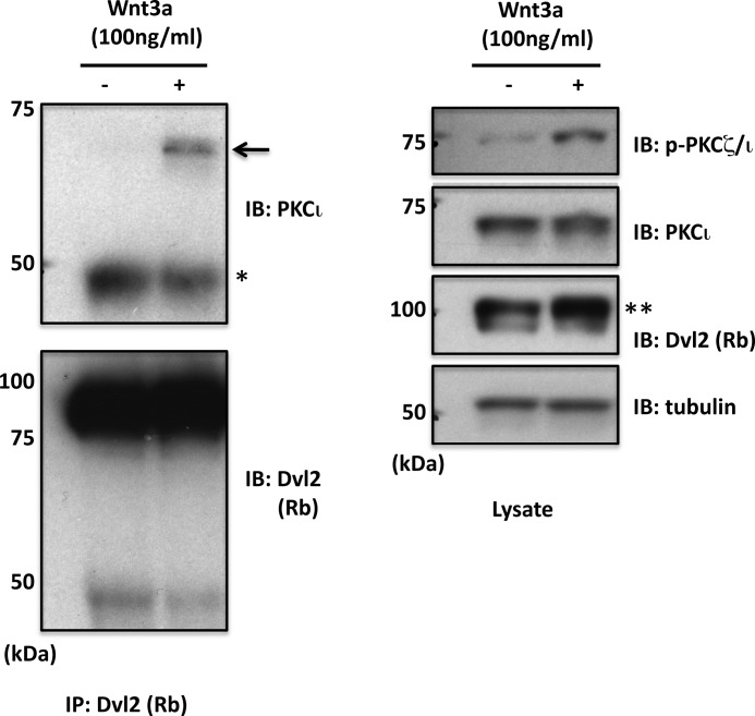
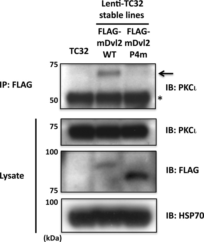
Previously we reported that Wnt3a-dependent neurite outgrowth in Ewing sarcoma family tumor cell lines was mediated by Frizzled3, Dishevelled (Dvl), and c-Jun N-terminal kinase (Endo, Y., Beauchamp, E., Woods, D., Taylor, W. G., Toretsky, J. A., Uren, A., and Rubin, J. S. (2008) Mol. Cell. Biol. 28, 2368-2379). Subsequently, we observed that Dvl2/3 phosphorylation correlated with neurite outgrowth and that casein kinase 1δ, one of the enzymes that mediate Wnt3a-dependent Dvl phosphorylation, was required for neurite extension (Greer, Y. E., and Rubin, J. S. (2011) J. Cell Biol. 192, 993-1004). However, the functional relevance of Dvl phosphorylation in neurite outgrowth was not established. Dvl1 has been shown by others to be important for axon specification in hippocampal neurons via an interaction with atypical PKCζ, but the role of Dvl phosphorylation was not evaluated. Here we report that Ewing sarcoma family tumor cells express PKCι but not PKCζ. Wnt3a stimulated PKCι activation and caused a punctate distribution of pPKCι in the neurites and cytoplasm, with a particularly intense signal at the centrosome. Knockdown of PKCι expression with siRNA reagents blocked neurite formation in response to Wnt3a. Aurothiomalate, a specific inhibitor of PKCι/Par6 binding, also suppressed neurite extension. Wnt3a enhanced the co-immunoprecipitation of endogenous PKCι and Dvl2. Although FLAG-tagged wild-type Dvl2 immunoprecipitated with PKCι, a phosphorylation-deficient Dvl2 derivative did not. This derivative also was unable to rescue neurite outgrowth when endogenous Dvl2/3 was suppressed by siRNA (González-Sancho, J. M., Greer, Y. E., Abrahams, C. L., Takigawa, Y., Baljinnyam, B., Lee, K. H., Lee, K. S., Rubin, J. S., and Brown, A. M. (2013) J. Biol. Chem. 288, 9428-9437). Taken together, these results suggest that site-specific Dvl2 phosphorylation is required for Dvl2 association with PKCι. This interaction is likely to be one of the mechanisms essential for Wnt3a-dependent neurite outgrowth.


Figures

FIGURE 1.
FIGURE 1.
PKCι is expressed by TC-32 cells and phosphorylated in response to Wnt3a. A, immunoblot analysis of PKCι and PKCζ in HEK293 and TC-32 cell lysates. PKCι was detected in TC-32 cells, but contradictory results were obtained for PKCζ with two different antibodies. B, combination of immunoblot analysis and isoform-specific siRNA indicated that PKCι, but not PKCζ, is expressed by TC-32 cells. PKCζ antiserum from Santa Cruz reacts with both PKCζ and PKCι. Note that knockdown in HEK293 confirmed the specificity of the PKCι and PKCζ siRNA reagents. C, time course of PKCι phosphorylation stimulated by Wnt3a (antibody recognizes Thr412 in PKCι and Thr410 in PKCζ). D, quantitative results of Wnt3a-dependent PKCι phosphorylation. The band intensity of phospho-PKCι was normalized to loading control (HSP70 or tubulin). The graph shows average values ± S.D. of five independent experiments. *, p < 0.05; **, p < 0.01 compared with zero time point. Similar results were obtained when data were normalized to total PKCι. However, because of fluctuation in total PKCι levels, the statistical significance was evident only at the 3-h time point. IB, immunoblot.
FIGURE 2.
FIGURE 2.
Immunofluorescent analysis of pPKCι and total PKCι in TC-32 cells with or without Wnt3a treatment. The cells were transfected with luciferase (Luc; A and C) or PKCι (B and D) siRNA reagents and 48 h later incubated with serum-free culture medium ± Wnt3a (100 ng/ml) for 1 h, fixed in formaldehyde, and then stained for phalloidin, DNA (DAPI), and pPKCι (A and B) or total PKCι (C and D). A scale bar is indicated. Ctl., control.
FIGURE 3.
FIGURE 3.
Co-localization of pPKCι and total PKCι with the centrosomal marker pericentrin. A, confocal micrographs of TC-32 cells treated in the absence or presence of Wnt3a (100 ng/ml) for the indicated times and subsequently stained with DAPI and antibodies to pPKCζ/ι and pericentrin. Arrows point to centrosomes. B, percentage of cells in different treatment groups with pericentrosomal distribution of pPKCι. The cumulative number (n) of cells analyzed in more than three experiments is indicated. Wnt3a treatment for 1 and 3 h increased the percentage of cells with pericentrosomal distribution of pPKCι. p < 0.001 for both time points compared with negative control, Fisher's exact test in SigmaPlot (Systat Software Inc., San Jose, CA). C and D, the specificity of pPKCι (C) and total PKCι (D) immunofluorescent staining was tested by comparing signal obtained after treatment with luciferase (Luc, upper rows) versus PKCι (lower rows) siRNA as described in the Fig. 2 legend (Wnt3a was not used in this experiment). The cells were fixed in methanol. A scale bar is indicated. Ctl., control.
FIGURE 4.
FIGURE 4.
Knockdown of PKCι expression with siRNA blocked Wnt3a-dependent neurite outgrowth. A, representative confocal micrographs, PKCι siRNA versus luciferase siRNA control. TC-32 cells were treated with the indicated siRNA reagents and 48 h later incubated for 3 h with serum-free culture medium ± Wnt3a (100 ng/ml). B, quantitative summary of the percentage of cells with a neurite at least one cell diameter in length, normalized to luciferase siRNA control without Wnt3a treatment (typically ∼10% cells have a long neurite in this control). The bar graph shows the results of three independent experiments (mean ± S.D.). C, immunoblot confirmed knockdown of endogenous PKCι protein. Ctl., control; IB, immunoblot; Luc, luciferase.
FIGURE 5.
FIGURE 5.
ATM blocked Wnt3a-dependent neurite outgrowth. A, TC-32 cells were preincubated for 30 min with the indicated concentrations of ATM in serum-free culture fluid and then treated with Wnt3a (0 or 100 ng/ml) for 3 h. Quantification of neurite outgrowth was performed as described in the Fig. 4 legend. The bar graph shows the results of three independent experiments (mean ± S.D.). B, representative confocal micrographs of cells from the different treatment groups. Ctl., control.
FIGURE 6.
FIGURE 6.
Co-immunoprecipitation of endogenous PKCι and Dvl2 following Wnt3a treatment. TC-32 cells were incubated ± Wnt3a for 3 h, and cell lysates were prepared for immunoprecipitation with Dvl2 rabbit polyclonal antiserum. Pelleted protein and cell lysates were immunoblotted as indicated. An arrow points to immunoprecipitated PKCι. *, IgG heavy chain. **, note increased relative intensity of slower migrating Dvl2 band. IB, immunoblot; IP, immunoprecipitation.
FIGURE 7.
FIGURE 7.
Co-immunoprecipitation of FLAG-tagged Dvl2 derivatives and endogenous PKCι. FLAG-tagged mouse Dvl2 WT or a Wnt-induced phosphorylation mutant containing four alanine substitutions (Dvl2 P4m) was stably expressed in TC-32 using a lentiviral vector. The cells were lysed, and the proteins were precipitated with FLAG antibody. Pellets and cell lysates were immunoblotted as indicated. An arrow points to immunoprecipitated PKCι. *, IgG heavy chain. IB, immunoblot; IP, immunoprecipitation.

References

    1. Logan C. Y., Nusse R. (2004) The Wnt signaling pathway in development and disease. Annu. Rev. Cell Dev. Biol. 20, 781–810 - PubMed
    1. Fuerer C., Nusse R., Ten Berge D. (2008) Wnt signalling in development and disease. Max Delbruck Center for Molecular Medicine meeting on Wnt signaling in Development and Disease. EMBO Rep. 9, 134–138 - PMC - PubMed
    1. Malaterre J., Ramsay R. G., Mantamadiotis T. (2007) Wnt-Frizzled signalling and the many paths to neural development and adult brain homeostasis. Front. Biosci. 12, 492–506 - PubMed
    1. Montcouquiol M., Crenshaw E. B., 3rd, Kelley M. W. (2006) Noncanonical Wnt signaling and neural polarity. Annu. Rev. Neurosci. 29, 363–386 - PubMed
    1. Bovolenta P., Rodriguez J., Esteve P. (2006) Frizzled/RYK mediated signalling in axon guidance. Development 133, 4399–4408 - PubMed

Publication types

MeSH terms

LinkOut - more resources