

Pseudoaneurysm of the hepatic artery, a rare complication of an amebic liver abscess
- PMID: 23396987
- PMCID: PMC3553300
- DOI: 10.1016/j.jus.2009.02.003
Pseudoaneurysm of the hepatic artery, a rare complication of an amebic liver abscess
Abstract
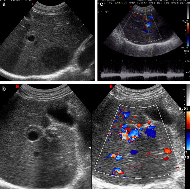
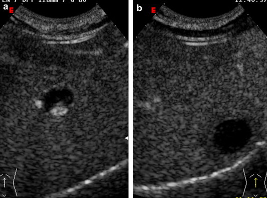
Hepatic artery pseudoaneurysm is a rare complication of amebic or pyogenic liver abscesses, and it is generally diagnosed because of hemobilia due to rupture of the aneurysm into the biliary tract. The authors describe a case of vascular complication in a patient affected by amebic liver abscess. Pseudoaneurysm was diagnosed and resolved without hemobilia.
SommarioGli pseudoaneurismi dell'arteria epatica sono una complicanza molto rara degli ascessi epatici amebici e da piogeni e la maggior parte vengono diagnosticati perché si complicano con emobilia.Gli autori descrivono un caso di complicanza vascolare in un paziente affetto da ascesso amebico del fegato diagnosticato e risolto senza la rottura nelle vie biliari.
Keywords: Abscess; Amebiasis; Pseudoaneurysm.
Figures


Similar articles
-
A case of amebic liver abscess complicated by hemobilia due to rupture of hepatic artery aneurysm.Hepatogastroenterology. 2002 Mar-Apr;49(44):375-8. Hepatogastroenterology. 2002. PMID: 11995454
-
Hepatic Abscess in a Returning Traveler with Crohn's Disease: Differentiating Amebic from Pyogenic Liver Abscess.Case Rep Med. 2018 May 29;2018:9593865. doi: 10.1155/2018/9593865. eCollection 2018. Case Rep Med. 2018. PMID: 30002680 Free PMC article.
-
Hepatic artery pseudoaneurysm associated with amebic liver abscess presenting as upper GI hemorrhage.Am J Gastroenterol. 1997 Aug;92(8):1391-3. Am J Gastroenterol. 1997. PMID: 9260822
-
Thoracic amebiasis.Clin Chest Med. 2002 Jun;23(2):479-92. doi: 10.1016/s0272-5231(01)00008-9. Clin Chest Med. 2002. PMID: 12092041 Review.
-
Surgical management of hepatic abscesses.World J Surg. 1990 Jul-Aug;14(4):498-504. doi: 10.1007/BF01658675. World J Surg. 1990. PMID: 2200212 Review.
Cited by
-
Successful peroral cholangioscopic extraction of migrated endovascular coils into the bile ducts 2 years following right hepatic artery pseudoaneurysm endovascular treatment.VideoGIE. 2024 Apr 20;9(8):379-381. doi: 10.1016/j.vgie.2024.04.004. eCollection 2024 Aug. VideoGIE. 2024. PMID: 39233839 Free PMC article. No abstract available.
-
Case Report: Spontaneous Resolution of Intracavitary Hepatic Artery Pseudoaneurysm Caused by Amebic Liver Abscess following Percutaneous Drainage.Am J Trop Med Hyg. 2019 Jul;101(1):157-159. doi: 10.4269/ajtmh.19-0103. Am J Trop Med Hyg. 2019. PMID: 31162010 Free PMC article.
-
Liver Abscess Caused by Klebsiella oxytoca with Hepatic Artery Pseudoaneurysm: A Case Report.Taehan Yongsang Uihakhoe Chi. 2020 Nov;81(6):1448-1452. doi: 10.3348/jksr.2019.0194. Epub 2020 Sep 1. Taehan Yongsang Uihakhoe Chi. 2020. PMID: 36237704 Free PMC article.
-
Radiological management of multiple hepatic artery pseudoaneurysms associated with cholangitic abscesses.Indian J Radiol Imaging. 2016 Jan-Mar;26(1):99-102. doi: 10.4103/0971-3026.178353. Indian J Radiol Imaging. 2016. PMID: 27081232 Free PMC article.
References
-
- Stanley S.L., Jr. Amoebiasis. Lancet. 2003;361:1025–1034. - PubMed
-
- Wells C.D., Arguedas M. Amoebic liver abscess. South Med J. 2004;97:673–682. - PubMed
-
- Krishnan K., Badarinath S., Bhusnurmath S.R. Vascular complications of hepatic amoebiasis: a retrospective study. Indian J Pathol Microbiol. 1986;29:293–296. - PubMed
-
- Sodhi K.S., Ojili V., Sakhuja V., Khandelwal N. Hepatic and inferior vena caval thrombosis: vascular complication of amebic liver abscess. J Emerg Med. 2008;34:155–157. - PubMed
-
- Yanagisawa M., Kaneko M., Aizawa T., Michimata T., Takagi H., Mori M. A case of amebic abscess complicated by hemobilia due to rupture of hepatic artery aneurysm. Hepatogastroenterology. 2002;49:375–378. - PubMed
LinkOut - more resources
Full Text Sources