

Contrast-enhanced sonography in blunt scrotal trauma()
- PMID: 23396988
- PMCID: PMC3558072
- DOI: 10.1016/j.jus.2011.09.003
Contrast-enhanced sonography in blunt scrotal trauma()
Abstract
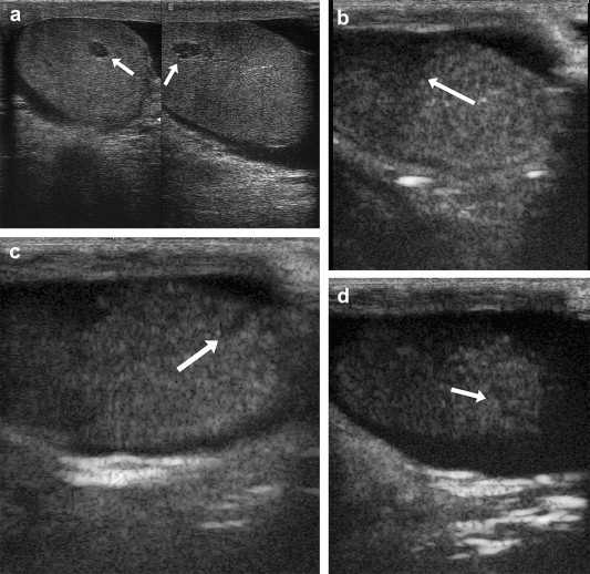
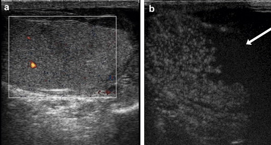
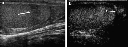
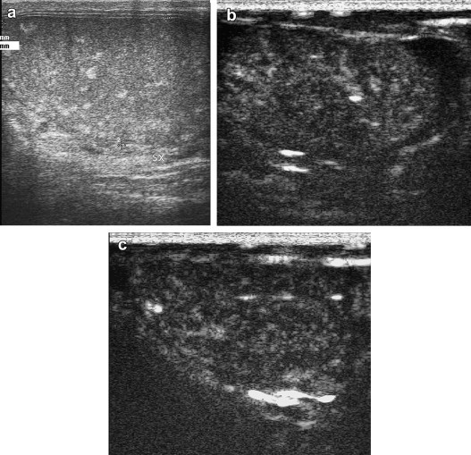
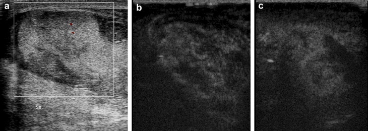
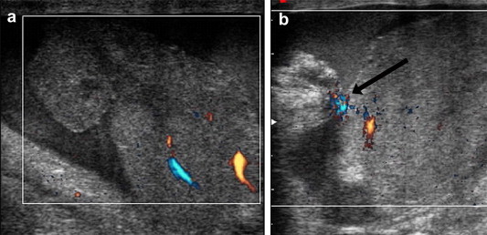
The scope of this study was to determine whether contrast-enhanced ultrasonography (CEUS), compared with basic US, can increase diagnostic confidence and provide relevant information on blunt scrotal trauma. Over a period of 75 months we examined 40 patients seen consecutively for blunt scrotal trauma using high-resolution US, color-power Doppler, low mechanical index CEUS, and power Doppler after IV administration of contrast medium (SonoVue(®)). In the 24 cases that were positive, concordance between basal US and CEUS findings was grade 0 (absent) in 4 cases, grade 1 (low) in 3, grade 2 (moderate) in 8, and grade 3 (high) in 9. The relevance of the additional information provided by CEUS was classified as follows: high in 4/40 (10%), moderate 7/40 (17,5%), low 13/40 (32,5%), none in 14/40 (35%). Our findings demonstrate that CEUS is appreciably more sensitive in detecting damage caused by blunt scrotal trauma, particularly small lesions. It is also useful for differential diagnosis and marginalization of corpuscular fluid collections, fractures, and above all ruptures, which require immediate surgery. In our series 2 out of 3 (67%) patients with testicular rupture were diagnosed only by CEUS. We feel that the use of CEUS can significantly improve diagnostic confidence in cases of closed scrotal trauma although these conclusions need to be confirmed in larger case series.
SommarioScopo del nostro lavoro è stato di valutare l’eventuale maggiore confidenza diagnostica e contenuto informativo dell’ecografia con mezzo di contrasto e.v. (CEUS) nel trauma scrotale chiuso rispetto all’indagine ecografica (US) di base. Nell’arco di 75 mesi abbiamo esaminato 40 pazienti consecutivi con trauma scrotale chiuso, utilizzando US ad alta risoluzione, color-power-Doppler basale, ecocontrastografia a basso indice meccanico, power Doppler dopo mdc e.v. Il mdc usato è stato il SonoVue. Nei 24 casi positivi, la concordanza tra US basale e CEUS è stata di grado 0 (assente) in 4 casi, di grado 1 (bassa) in 3, di grado 2 (medio) in 8, di grado 3 (elevato) in 9. La rilevanza del contenuto informativo aggiuntivo della CEUS veniva ritenuta: elevata in 4/40 (10%), media 7/40 (17,5%), bassa 13/40 (32,5%), assente in 14/40 (35%). I nostri risultati mostrano che la CEUS migliora sensibilmente la detezione dei segni di trauma rispetto l’US basale, specialmente nelle piccole lesioni. Essa è inoltre importante nella diagnosi differenziale e marginalizzazione delle raccolte fluide corpuscolate, nelle fratture e, di grande evidenza, nelle rotture, che impongono l’immediato intervento chirurgico: nella nostra casistica 2 casi su 3 di rotture (67%) si sono resi evidenti soltanto alla CEUS. Riteniamo che l’uso della CEUS possa aumentare significativamente la confidenza nella diagnostica del trauma scrotale chiuso, ma sono necessarie conferme da casistiche più sostanziose.
Keywords: Acute scrotum; Contrast media; Contrast-enhanced sonography; Scrotal trauma; Sonography.
Figures
Similar articles
-
Diagnostic value of contrast-enhanced ultrasound (CEUS) and comparison with color Doppler ultrasound and magnetic resonance in a case of scrotal trauma.J Ultrasound. 2020 Jun;23(2):189-194. doi: 10.1007/s40477-019-00389-y. Epub 2019 Jun 5. J Ultrasound. 2020. PMID: 31168706 Free PMC article.
-
Role of contrast enhanced ultrasound in acute scrotal diseases.Eur Radiol. 2011 Sep;21(9):1831-40. doi: 10.1007/s00330-010-2039-5. Epub 2011 Jun 2. Eur Radiol. 2011. PMID: 21633826
-
Accuracy of contrast-enhanced ultrasound (CEUS) in the identification and characterization of traumatic solid organ lesions in children: a retrospective comparison with baseline US and CE-MDCT.Radiol Med. 2015 Nov;120(11):989-1001. doi: 10.1007/s11547-015-0535-z. Epub 2015 Mar 31. Radiol Med. 2015. PMID: 25822953
-
Use of contrast enhanced ultrasound in testicular diseases: A comprehensive review.Andrology. 2021 Sep;9(5):1369-1382. doi: 10.1111/andr.13057. Epub 2021 Jun 11. Andrology. 2021. PMID: 34043256 Free PMC article. Review.
-
Sonography of pediatric blunt scrotal trauma: what the pediatric urologist wants to know.Pediatr Radiol. 2016 Jun;46(7):1049-58. doi: 10.1007/s00247-016-3600-4. Epub 2016 Apr 25. Pediatr Radiol. 2016. PMID: 27112160 Review.
Cited by
-
Diagnosis and management of testicular rupture after blunt scrotal trauma: a literature review.Int Urol Nephrol. 2016 Dec;48(12):1967-1976. doi: 10.1007/s11255-016-1402-0. Epub 2016 Aug 27. Int Urol Nephrol. 2016. PMID: 27567912 Review.
-
The role of contrast-enhanced ultrasound (CEUS) in the evaluation of scrotal trauma: a review.Insights Imaging. 2020 May 19;11(1):68. doi: 10.1186/s13244-020-00874-7. Insights Imaging. 2020. PMID: 32430792 Free PMC article. Review.
-
Contrast-enhanced ultrasound of blunt abdominal trauma in children.Pediatr Radiol. 2021 Nov;51(12):2253-2269. doi: 10.1007/s00247-020-04869-w. Epub 2021 May 12. Pediatr Radiol. 2021. PMID: 33978795 Review.
-
Can ultrasound help to manage patients with scrotal trauma?Ultrasound. 2014 Nov;22(4):205-12. doi: 10.1177/1742271X14545911. Epub 2014 Jul 30. Ultrasound. 2014. PMID: 27433221 Free PMC article. Review.
-
Imaging in scrotal trauma: a European Society of Urogenital Radiology Scrotal and Penile Imaging Working Group (ESUR-SPIWG) position statement.Eur Radiol. 2021 Jul;31(7):4918-4928. doi: 10.1007/s00330-020-07631-w. Epub 2021 Jan 15. Eur Radiol. 2021. PMID: 33449189
References
-
- Bhatt S., Dogra V.S. Role of US in testicular and scrotal trauma. RadioGraphics. 2008;28:1617–1629. - PubMed
-
- Deurdulian C., Mittelstaedt C.A., Chong W.K., Fielding J.R. US of acute scrotal trauma: optimal technique, imaging findings, and management. Radiographics. 2007;27:357–369. - PubMed
-
- Buckley J.C., McAninch J.W. Use of ultrasonography for the diagnosis of testicular injuries in blunt scrotal trauma. J Urol. 2006;175:175–178. - PubMed
-
- Cass A.S., Luxenberg M. Testicular injuries. Urology. 1991;37:528–530. - PubMed
-
- Gross M. Rupture of the testicle: the importance of early surgical treatment. J Urol. 1969;101:196–197. - PubMed
LinkOut - more resources
Full Text Sources