

Cysts of the canal of Nuck: ultrasound and magnetic resonance imaging findings
- PMID: 23397010
- PMCID: PMC3567455
- DOI: 10.1016/j.jus.2009.05.002
Cysts of the canal of Nuck: ultrasound and magnetic resonance imaging findings
Abstract
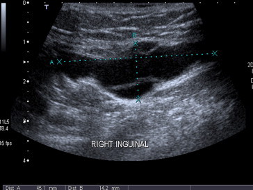
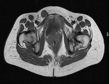
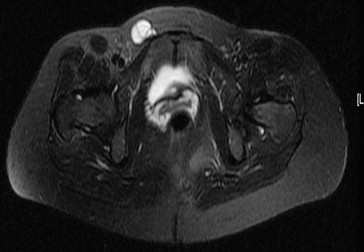
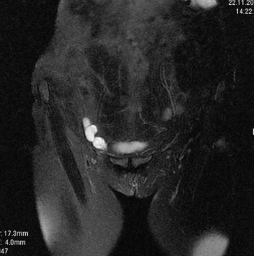
Cyst of the canal of Nuck is a rare cause of inguinal swelling in woman. We report a case of a cyst of the canal of Nuck in which sonography showed a tubular cystic structure with internal septae localized within the inguinal canal. Magnetic resonance examination demonstrated that the mass was hypointense on T1-weighted and hyperintense on T2-weighted series and that there were fine hypointense septae inside the mass on the T2-weighted sequence. Diagnosis of cyst of the canal of Nuck was confirmed by surgery and subsequent histopathologic evaluation.
SommarioLa cisti del canale di Nuck è una rara causa di tumefazione inguinale nelle donne. Riportiamo un caso di cisti del canale di Nuck in cui l'ecografia aveva evidenziato una struttura tubulare cistica caratterizzata da setti interni, localizzata nel contesto del canale inguinale. L'esame di RM aveva confermato la natura cistica della lesione, che appariva pertanto ipointensa in T1 e iperintensa con fini sepimentazioni ipointense nelle sequenze T2 pesate. La diagnosi di cisti del canale di Nuck è stata successivamente confermata all'intervento chirurgico e dall'esame isto-patologico.
Keywords: Canal of Nuck; Cyst; Groin; Hydrocele; Magnetic resonance imaging; Sonography.
Figures
References
-
- Anderson C.C., Broadie T.A., Mackey J.E., Kopecky K.K. Hydrocele of the canal of Nuck: ultrasound appearance. Am Surg. 1995;61:959–961. - PubMed
-
- Ihekwaba F.N. Hydrocele in the female. J R Coll Surg Edinb. 1981;26:91–93. - PubMed
-
- Kucera P.R., Glazer J. Hydrocele of the canal of Nuck: a report of 4 cases. J Reprod Med. 1985;30:439–442. - PubMed
-
- Walter H.S., Martin M. Female hydrocele (cyst of the canal of Nuck) J Ultrasound Med. 2004;23:429–432. - PubMed
-
- Park S.J., Lee H.K., Hong H.S. Hydrocele of the canal of Nuck in a girl: ultrasound and MR appearance. Br J Radiol. 2004;77:243–244. - PubMed
LinkOut - more resources
Full Text Sources