

Renal arteriovenous fistula simulating hydronephrosis: A case report
- PMID: 23397018
- PMCID: PMC3558108
- DOI: 10.1016/j.jus.2011.10.006
Renal arteriovenous fistula simulating hydronephrosis: A case report
Abstract
Introduction: Acquired renal arteriovenous fistulas (AVFs) include those that occur as a complication of renal biopsy.
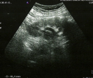
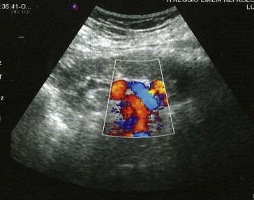
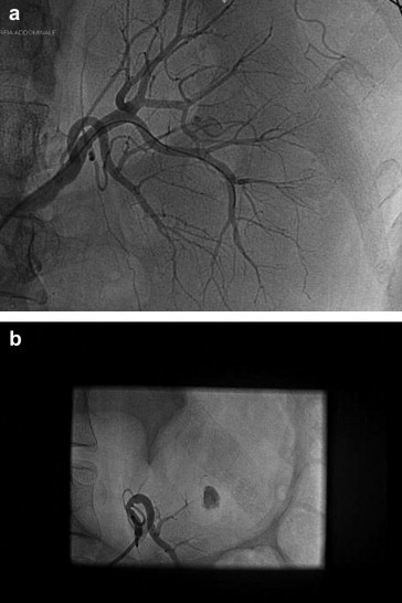
Case report: The authors report the case of a woman with recent-onset grade I hypertension, who was referred to our staff for sonographic studies of the kidneys and urinary tract. Laboratory data revealed microhematuria and proteinuria <0.5 g/24 h, and renal function was borderline (MDRD GFR 58 mL/min). Renal sonography of the left kidney revealed an anechoic, arboriform area at the level of the pelvis, which was suggestive of hydronephrosis. The color Doppler examination showed turbulent flow within the anechoic area, with high-velocity arterial flow and arterialization of the venous waveform at spectral analysis. Selective renal angiography later confirmed the presence of a middle renal AVF with pseudoaneurysm, which had been provoked by a renal biopsy performed over 10 years earlier in another center. Since the patient was currently in good health, the prescribed management consisted solely of close clinical and US follow-up.
Discussion: AV fistulas are among the most commonly diagnosed renovascular malformations. The case reported here underlines the importance of using color Doppler ultrasound when obstructive uropathy is suspected, especially in patients who have undergone renal biopsy.
Sommario INTRODUZIONE: Tra le fistole arterovenose (FAV) acquisite vi sono quelle che si formano quale complicanza dell’esecuzione di una biopsia renale. CASO CLINICO: Gli autori riportano il caso di una paziente giunta in ambulatorio di ecografia nefrologica per l’esecuzione di un’ecografia dei reni e delle vie urinarie per un’ipertensione sistolica di I grado di recente insorgenza. Negli esami di laboratorio era presente microematuria con proteinuria <0.5 g/24 ore. La funzionalità renale risultava ai limiti inferiori della norma (FG 58 mL/min secondo la formula MDRD). L’esame ecografico evidenziava una formazione anecogena “arboriforme” centropielica a livello del rene sinistro suggestiva per idronefrosi. Il color Doppler mostrava viceversa presenza di flusso turbolento all’interno dell’area anecogena stessa, con flusso arterioso elevato e arterializzazione del flusso venoso all’analisi spettrale. La successiva angiografia renale selettiva confermava il sospetto ecografico di FAV mediorenale sinistra con pseudoaneurisma, conseguente ad una biopsia renale effettuata più di 10 anni prima presso altro Centro. Essendo la paziente in pieno benessere veniva consigliato un più stretto follow-up clinico-ecografico. DISCUSSIONE: Tra le malformazioni vascolari renali di non rara osservazione sono le FAV. Il caso descritto ribadisce l’importanza dell’impiego dell’eco color Doppler (ECD) nel sospetto di uropatia ostruttiva, per diagnosticare una FAV, specialmente nei pazienti già sottoposti a biopsia renale.
Keywords: Hydronephrosis; Pseudoaneurysm; Renal arteriovenous fistula; Renal arteriovenous malformations.
Figures

References
-
- Kopecna L., Mach V., Prochazka J. Arteriovenous fistula as a complication of renal biopsy. Bratisl Lek Listy. 2005;106(6–7):218–220. - PubMed
-
- Rivera M., Villacorta J., Jimenez-Alvaro S., Quereda C. Asymptomatic large extracapsular renal pseudoaneurysm following kidney transplant biopsy. Am J Kidney Dis. 2011;57(1):175–178. - PubMed
-
- Kember P.G., Peck R.J. Renal arteriovenous malformation mimicking hydronephrosis. J Clin Ultrasound. 1998;26:95–97. - PubMed
-
- Li J.C., Cai S., Jiang Y.X., Dai Q., Zhang J.X., Wang Y.Q. Diagnostic criteria for locating acquired arteriovenous fistulas with color Doppler sonography. J Clin Ultrasound. 2002;30(6):336–342. - PubMed
LinkOut - more resources
Full Text Sources