

Clues to the etiology of bile duct injury in biliary atresia
- PMID: 23397531
- PMCID: PMC3827890
- DOI: 10.1055/s-0032-1329899
Clues to the etiology of bile duct injury in biliary atresia
Abstract
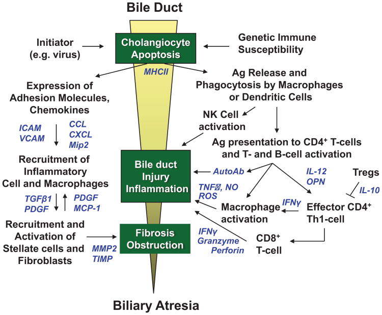
Biliary atresia (BA) is an infantile obstructive cholangiopathy of unknown etiology with suboptimal therapy, which is responsible for 40 to 50% of all pediatric liver transplants. Although the etiology of bile duct injury in BA in unknown, it is postulated that a pre- or perinatal viral infection initiates cholangiocyte apoptosis and release of antigens that trigger a Th1 immune response that leads to further bile duct injury, inflammation, and obstructive fibrosis. Humoral immunity and activation of the innate immune system may also play key roles in this process. Moreover, recent investigations from the murine BA model and human data suggest that regulatory T cells and genetic susceptibility factors may orchestrate autoimmune mechanisms. What controls the coordination of these events, why the disease only occurs in the first few months of life, and why a minority of infants with perinatal viral infections develop BA are remaining questions to be answered.
Thieme Medical Publishers 333 Seventh Avenue, New York, NY 10001, USA.
Figures
References
-
- Mack CL, Sokol RJ. Unraveling the pathogenesis and etiology of biliary atresia. Pediatr Res. 2005;57(5 Pt 2):87R–94R. - PubMed
-
- Sokol RJ, Mack C, Narkewicz MR, Karrer FM. Pathogenesis and outcome of biliary atresia: current concepts. J Pediatr Gastroenterol Nutr. 2003;37(1):4–21. - PubMed
-
- Hartley JL, Davenport M, Kelly DA. Biliary atresia. Lancet. 2009;374(9702):1704–1713. - PubMed
-
- Petersen C. Pathogenesis and treatment opportunities for biliary atresia. Clin Liver Dis. 2006;10(1):73–88,vivi. - PubMed
Publication types
MeSH terms
Grants and funding
LinkOut - more resources
Full Text Sources
Other Literature Sources
Medical
Molecular Biology Databases
