. 2012:763:260-80.
doi: 10.1007/978-1-4614-4711-5_13.

Gap junctions and blood-tissue barriers

Affiliations

Gap junctions and blood-tissue barriers

Michelle W M Li et al. Adv Exp Med Biol. 2012.

Abstract

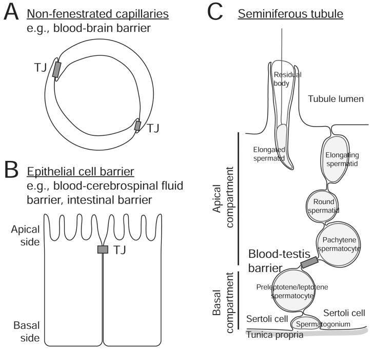
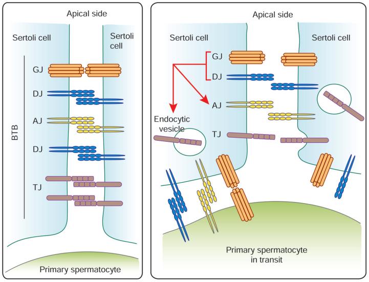
Gap junction is a cell-cell communication junction type found in virtually all mammalian epithelia and endothelia and provides the necessary "signals" to coordinate physiological events to maintain the homeostasis of an epithelium and/or endothelium under normal physiological condition and following changes in the cellular environment (e.g., stimuli from stress, growth, development, inflammation, infection). Recent studies have illustrated the significance of this junction type in the maintenance of different blood-tissue barriers, most notably the blood-brain barrier and blood-testis barrier, which are dynamic ultrastructures, undergoing restructuring in response to stimuli from the environment. In this chapter, we highlight and summarize the latest findings in the field regarding how changes at the gap junction, such as the result of a knock-out, knock-down, knock-in, or gap junction inhibition and/or its activation via the use of inhibitors and/or activators, would affect the integrity or permeability of the blood-tissue barriers. These findings illustrate that much research is needed to delineate the role of gap junction in the blood-tissue barriers, most notably its likely physiological role in mediating or regulating the transport of therapeutic drugs across the blood-tissue barriers.


Figures

Figure 1
Figure 1
Schematic illustrations of the structure and organization of GJ. A) A connexin consists of four transmembrane domains, two extracellular loops and one intracellular loop. The variability of connexins lies mostly on the C-terminal tail that comes in different length and sequence and carries sites for phosphorylation and binding of interacting partners. B) Six connexins constitute a functional connexon. A connexon can be made up of the same type of connexins (homomeric) or of different types (heteromeric). An uncoupled connexon can also be called a hemichannel. C) GJ channel is formed between two compatible connexons on adjacent to that create a functional communication channel. The interaction of connexons could be homotypic or heterotypic, depending on the compatibility of individual connexins in a connexon.
Figure 2
Figure 2
This figure illustrates the major morphological features of various types of blood-tissue barriers. A) Blood-tissue barriers, including blood-brain barrier, could be constituted by TJ formed between endothelial vascular cells in nonfenestrated capillaries. B) Blood-tissue barriers could also be formed by epithelial cells. At the blood-cerebrospinal fluid barrier, the blood vessel is fenestrated (without TJ) and TJs formed at the apical region of adjacent choroid plexus epithelial cells constitute the barrier. C) The blood-testis barrier is located in the seminiferous epithelium of the seminiferous tubule, which is formed near the basal region of adjacent Sertoli cells. Different junction complexes have been identified at this site, including TJ, basal ES (an atypical AJ), desmosome-like junction and GJ. The blood-testis barrier also segregates the seminiferous epithelium into the basal and apical (or adluminal) compartment, so that meiosis and the entire events of postmeiotic germ cell development (i.e., spermiogenesis) take place behind this immunological barrier in a specialized microenvironment.
Figure 3
Figure 3
Schematic illustration of the roles of GJ and desmosome at the blood-testis barrier. The blood-testis barrier (BTB) remains intact at most stages of the seminiferous epithelial cycle and consists of coexisting TJ, AJ, GJ and desmosome-like junction (DJ) (left panel). Primary spermatocytes migrate across the BTB at Stage VIII of the seminiferous epithelial cycle in the rat testis (right panel). The AJ, DJ and GJ formed between Sertoli cells are likely replaced by those between Sertoli cell and spermatocyte since it is now known that many of AJ, DJ and GJ proteins are also found in germ cells, such as spermatocytes.- A reduction of GJ and DJ between adjacent Sertoli cells would destabilize the BTB, inducing its disruption. This involves a decline in the steady-state levels of TJ and AJ proteins at the Sertoli cell surface, which is partly mediated by an increase in endocytosis of junction proteins. The net result is an increase in the permeability at the apical region of the translocating spermatocytes to facilitate their transit at the BTB. However, “new” TJ, AJ, DJ and GJ are formed behind the spermatocytes in transit before the “old” junctions are being disrupted, so that the immunological barrier can be maintained.

References

    1. Alberts B, Johnson A, Lewis J, et al. Molecular Biology of the Cell. 4 th ed Garland Science; New York: 2002.
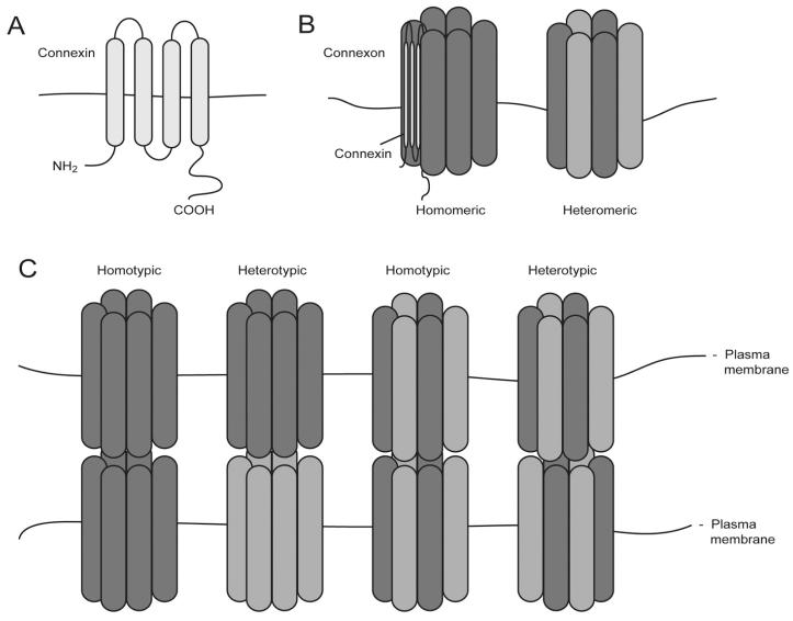
    1. Meşe G, Richard G, White TW. Gap junctions: basic structure and function. J Invest Dermatol. 2007;127:2516–2524. - PubMed
    1. Dbouk HA, Mroue RM, El-Sabban ME, et al. Connexins: a myriad of functions extending beyond assembly of gap junction channels. Cell Commun Signal. 2009;7:4. - PMC - PubMed
    1. Sáez JC, Berthoud VM, Brañes MC, et al. Plasma membrane channels formed by connexins: their regulation and functions. Physiol Rev. 2003;83:1359–1400. - PubMed
    1. Shestopalova VI, Panchinc Y. Pannexins and gap junction protein diversity. Cell Mol Life Sci. 2008;65:376–394. - PMC - PubMed

Publication types

MeSH terms

LinkOut - more resources