Review
. 2013 Nov 1;19(13):1539-605.
doi: 10.1089/ars.2012.4599. Epub 2013 Mar 28.

Thioredoxins, glutaredoxins, and peroxiredoxins--molecular mechanisms and health significance: from cofactors to antioxidants to redox signaling

Affiliations
Review

Thioredoxins, glutaredoxins, and peroxiredoxins--molecular mechanisms and health significance: from cofactors to antioxidants to redox signaling

Eva-Maria Hanschmann et al. Antioxid Redox Signal. .

Abstract

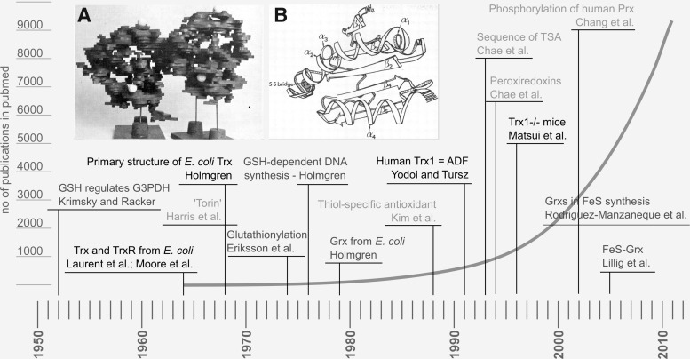
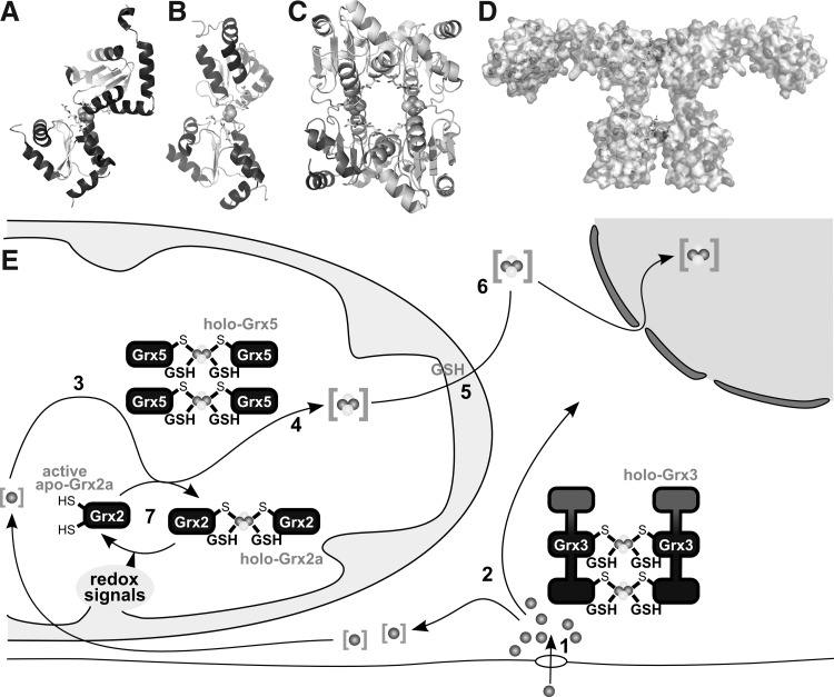
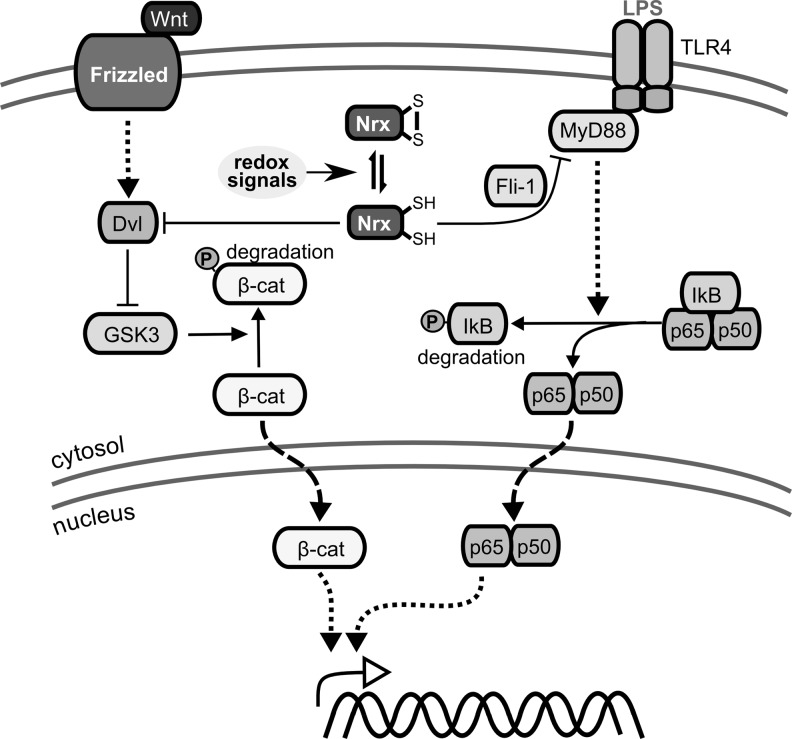
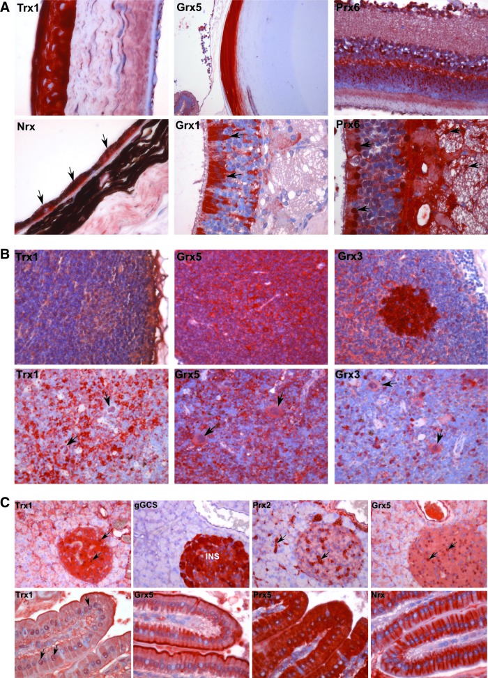
Thioredoxins (Trxs), glutaredoxins (Grxs), and peroxiredoxins (Prxs) have been characterized as electron donors, guards of the intracellular redox state, and "antioxidants". Today, these redox catalysts are increasingly recognized for their specific role in redox signaling. The number of publications published on the functions of these proteins continues to increase exponentially. The field is experiencing an exciting transformation, from looking at a general redox homeostasis and the pathological oxidative stress model to realizing redox changes as a part of localized, rapid, specific, and reversible redox-regulated signaling events. This review summarizes the almost 50 years of research on these proteins, focusing primarily on data from vertebrates and mammals. The role of Trx fold proteins in redox signaling is discussed by looking at reaction mechanisms, reversible oxidative post-translational modifications of proteins, and characterized interaction partners. On the basis of this analysis, the specific regulatory functions are exemplified for the cellular processes of apoptosis, proliferation, and iron metabolism. The importance of Trxs, Grxs, and Prxs for human health is addressed in the second part of this review, that is, their potential impact and functions in different cell types, tissues, and various pathological conditions.


Figures

FIG. 1.
FIG. 1.
A brief history of “redoxin” research. The figure highlights some milestones of Trx, Grx, and Prx research and (in the background) the number of publications listed in pubmed with the query “Trx OR Grx OR thioltransferase OR Prx”. Black: Trx, dark gray: Grx, and light gray: Prx-related findings. Insets: (A) The first structure of Escherichia coli Trx at 4–5 Å resolution, photography of the balsa model (Söderberg et al. 1974) (723). (B) Drawing of the first high-resolution structure of E. coli Trx at 2.8 Å (Holmgren et al. 1975) (293). The work by Krimsky and Racker in 1952 (408) on GSH and glyceralaldehyde-3-phosphate dehydrogenase did not decipher the redox nature of this interaction, but first emphasized the regulatory functions of GSH. GSH, glutathione; Trx, thioredoxin; Grx, glutaredoxin; Prx, peroxiredoxin; ADF, adult T-cell leukemia-derived factor; TSA, thiol-specific antioxidant.
FIG. 2.
FIG. 2.
The Trx fold. (A) Schematic representation of the Trx fold, the asterisk marks the position of the proximal active site cysteinyl residue, helices are shown in dark, sheets in light gray. Bacterial Grxs, such as (B) E. coli Grx1 (PDB accession number: 1EGR), are the most basic representations of the fold. (C) Human Trx1 (PDB: 3TRX) contains an additional N-terminal sheet and helix. (D, E) The 2-Cys Prx1 is shown as monomer (D) and (E) decameric torin.
FIG. 3.
FIG. 3.
Electron flow from NADPH to substrates via the Trx and GSH/Grx systems. NADPH as the main electron source reduces the selenoprotein thioredoxin reductase (TrxR), which delivers electrons to Trx, which then reduces protein (P) disulfides. NADPH also donates electrons to glutathione reductase (GR), which reduces glutathione disulfide (GSSG), thereby generating two molecules of reduced GSH. Electrons can then be delivered to oxidized Grx, which either possesses an active site disulfide bridge due to reduction of protein disulfides or a glutathionylated N-terminal active site Cys from reducing a GSH-mixed disulfide.
FIG. 4.
FIG. 4.
Reaction mechanisms of Trx family proteins. (A) Trxs reduce protein disulfides via the dithiol mechanism, depending on both active site cysteines. The N-terminal active site Cys forms a covalently bound mixed disulfide intermediate (A 1), which is reduced by the C-terminal active site Cys, releasing the reduced protein (A 2). Oxidized Trx is reduced by TrxR in a similar reaction sequence (A 3–4). (B) Grxs also reduce protein disulfides via the dithiol mechanism, being reduced by two GSH molecules (B 1–4). In addition, they reduce glutathionylated proteins via the monothiol mechanism (B 5–4), only depending on the N-terminal active site Cys, that attacks the GSH moiety and forms a GSH-mixed disulfide intermediate (B 5), which is reduced by another GSH molecule (B 4). (C) During the reduction of H2O2 by Prxs, the redox-active, peroxidatic Cys (labeled p) is oxidized to sulfenic acid (C 1), which either forms an inter-(2-Cys Prxs) (C 2) or an intramolecular disulfide (atypical 2-Cys Prxs) (not shown) with the resolving Cys residue (labeled r), with both being reduced by Trx as outlined in (A) (C 3–4). 1-Cys Prxs lack an additional resolving cysteine and are reduced by GSH (not shown). In the presence of H2O2, the sulfenic acid can be further oxidized (“over-oxidized”) to sulfinic acid [5] and sulfonic acid [9]. Sulfinic acid-modified Prxs can be recovered by the ATP-dependent action of sulfiredoxin (Srx) [6–8]. For a detailed discussion, see section I.A.1. H2O2, hydrogen peroxide.
FIG. 5.
FIG. 5.
Mammalian Trxs, Grxs, and Prxs. Isoforms, subcellular localization, and confirmed interactions between the various redox proteins discussed in this review. The active site sequences and the classes of proteins, respectively, are indicated in white. C, cytosol; M, mitochondrium; N, nucleus; P, peroxisome; S, secreted. The secretory compartments, that is, endoplasmatic reticulum, Golgi apparatus, and lysosomes, were excluded for reasons of clarity; however, these compartments contain Trx family proteins; see Table 1.
FIG. 6.
FIG. 6.
Production and reactivity of reactive nitrogen, oxygen, and sulfur species. RNS (bio-)chemistry, left side: [1] Production of nitric oxide by nitric oxide synthase (NOS). [2–3] S-nitrosylation of protein thiols. [4] Trans-nitrosylation between protein thiols. [5] Reaction of nitric oxide with metals, for example, heme iron. [6] Nitric oxide reacts spontaneously with superoxide yielding peroxynitrite. [7] Reversible protonation of peroxynitrite to peroxynitrous acid. [8] Reduction of peroxynitrous acid by glutathione peroxidases (GPxs) or PRX. [9] Peroxynitrous acid reacts with protein thiolates, yielding protein sulfenic acids. [10] Spontaneous decomposition of peroxynitrous acid yielding nitrite anion. [11] Spontaneous decomposition of peroxynitrous acid to hydroxy radicals and NO2·. [12] Peroxynitrite can (metal catalyzed) lead to the nitration of, for instance, protein tyrosyl residues. [13] Peroxynitrite and carbon dioxide react spontaneously to nitrosoperoxycarbonate. [14] Spontaneous decay of nitrosoperoxycarbonate to carbonate radical anions and nitrite radicals. [15] Spontaneous decay of nitrosoperoxycarbonate to carbon dioxide and nitrate. [16] Nitration may also be initiated by NO2·. ROS (bio-)chemistry, bmiddle: [17] Production of superoxide by, for instance, mitochondrial complex I (CI), NADH oxidase (NOX), cyclooxygenases (COX), xanthine oxidase (XO), or cytochrome P450 enzymes (Cp450). [18] Superoxide is either reduced to H2O2 or oxidized to molecular oxygen (not shown) by superoxide dismutases (SOD). [19] H2O2 can be reduced to water by GPxs or PRX. [20] H2O2 may react directly with specific thiols, yielding sulfenic acids. [21] Sulfenic acids can react with other thiols, yielding disulfides. These disulfides are direct substrates of Trxs and Grxs (not depicted). [22] Sulfenic acids may be further irreversibly oxidized, for example, by H2O2, to sulfinic and sulfonic acids. [23] H2O2 may react with chloride anions, yielding hypochlorous acid. [24] The metal-catalyzed Fenton reaction yields hydroxyl anions and hydroxy radicals. [25] Hydroxy radicals remove hydrogen from volatile organic compounds, yielding water and alkyl radicals. [26–27] Alkyl radicals may react with molecular oxygen and other compounds, eventually resulting in the peroxidation, carbonylation, or cleavage of the organic molecules, for example, proteins. [28] Hypochloric acid may lead to the chlorination of organic compounds. RSS biochemistry, right side: [29–32] Hydrogen sulfide may be the product of cystathionine β-synthase [29, CBS], cystathionine γ-lyase [30, CSE], or via 3-mercaptopyruvate sulfurtransferase [31–32, MST]. [33] Hydrogen sulfide may react with thiols in the presence of an electron and hydrogen acceptor to persulfides. Modifications labeled with a light gray background are reversible and important in redox signaling, and modifications with a dark gray background are irreversible modifications; hence, “oxidative damage.” ROS, reactive oxygen species; RNS, reactive nitrogen species; RSS, reactive sulfur species.
FIG. 7.
FIG. 7.
Redox modifications at cysteinyl residues. Free thiol groups (R-SH) can be reversibly modified by ROS, leading to the formation of protein disulfides (R-S-S-R), which can be reduced by the Trx and Grx systems. Thiols can also be glutathionylated (R-S-SG) by oxidized glutathione (GSSG) or S-nitroso glutathione (GSNO). The de-glutathionylation is exclusively catalyzed by Grxs. GSNO or ·NO, in general, can lead to the nitrosylation of cysteinyl residues, which can be reversed by GSH or transferred to other thiols such as the active site of Trx1 (53) (trans-nitrosylation, not shown). Another modification, induced by peroxides, is the formation of sulfenic acid (R-SOH). In the presence of another free thiol, it can be modified to a protein disulfide. However, in the presence of excessive peroxides, it can be irreversibly over-oxidized to sulfinic (R-SO2H) and sulfonic acid (R-SO3H). *The reduction of sulfinic acids to sulfenic acids, catalyzed by Srxs, is specific for Prxs; in addition, Srxs have been reported to catalyze the de-glutathionylation of Prxs.
FIG. 8.
FIG. 8.
Trx, Txnip, and Grx in MAP kinase and NF-κB signaling. Txnip, whose expression is promoted by glucose via MondoA:MLX signaling and repressed by FOXO1a, was suggested to be a negative regulator of reduced Trx1. Left side: Trx and Grx as negative regulators of apoptosis signal-regulating kinase 1 (ASK1)–ASK1 is a mitogen-activated protein (MAP) kinase kinase kinase that signals downstream to the c-Jun N-terminal kinase (JNK) and the p38 MAP kinase pathways via MAP kinase kinases 3, 4, 6, and 7. Reduced Trx1 and Grx1 can bind to ASK1, leading to an inactive complex. Oxidation of Trx1 and/or Grx1 by various redox signals leads to dissociation of the complex and activation of ASK1. Moreover, the Trx1/ASK1 complex is targeted for ubiquitination and degradation. Right side: Redox regulation of NF-κB activation–the NF-κB subunit p50 contains a cysteine (Cys 62) in its DNA binding site that is susceptible to oxidation. After dissociation of the I-κB/NF-κB complex, which is not only promoted by phosphorylation of I-κB in response to a variety of signals but also inhibited by reduced Trx1, NF-κB is translocated to the nucleus. In the nucleus, reduction of Cys62 in the p50 subunit of NF-κB is necessary for binding of the transcription factor to its target site in the DNA. In the nucleus, Trx1, Grx1, and Nrx (not shown) have been reported to promote NF-κB binding to the κB site in the DNA. NF-κB, nuclear factor kappa B; Nrx, nucleoredoxin; Txnip, trx interacting protein.
FIG. 9.
FIG. 9.
[FeS]-Grxs in cellular iron metabolism. (A) Structure of the holo-Grx2 complex consisting of two monomers Grx2 (cartoon graphics), two GSH molecules (ball and stick model), and the [2Fe2S] cluster (calotte model), derived from PDB entry 2HT9 (348). (B, C) Structures of the holo-Grx5 complex depicted as dimer (B) and tetrameric holo complexes (C), derived from PDB entry 2WUL (350). (D) Hypothetical model of the dimeric Grx3 holo complex (271). (E) Iron taken up into the cell, simplified in [1], is shuttled through the cytosol, presumably involving Grx3 [2]. Inside mitochondria, iron is used, for instance, for the biogenesis of iron-sulfur clusters [3] on a scaffold protein and transferred to target apo-proteins [4] in a reaction that requires Grx5. The export of iron-sulfur clusters in a hitherto unknown form requires GSH [5]. This compound X is used by the cytosolic iron-sulfur cluster assembly machinery for the synthesis of cytosolic and nuclear FeS proteins [6]. [7] Grx2 is usually present in the enzymatically inactive FeS-bridged dimeric holo form. On redox signals, the FeS cluster dissociates, yielding active monomeric Grx2.
FIG. 10.
FIG. 10.
Nrx in Wnt/Dvl and Toll-like receptor 4 (TLR4) signaling. Nrx was shown to suppress the Wnt/β-catenin pathway, which is involved in embryonic development and cancer. Secreted Wnt proteins bind to receptors of the Frizzled family and activate a signaling cascade. This process involves the cytosolic dishevelled (Dvl) protein, which inhibits the glycogen synthase kinase-3 (GSK3)-containing destruction apparatus and thereby phosphorylation and degradation of beta-catenin (β-cat). β-cat translocates into the nucleus and activates the transcription of Wnt-regulated target genes. Reduced Nrx binds to Dvl and suppresses Wnt/β-catenin signaling, whereas via “redox signals” oxidized Nrx does not. Similarly, reduced Nrx can inhibit TLR4 signaling, which is essential for embryonic development and the innate immune response. Lipopolysaccharide (LPS) stimulates the oligomerization of TLR4, inducing the recruitment of signal transduction adaptor proteins, such as myeloid differentiation primary response protein (MyD88). MyD88 activates a cascade of IKK and MAP kinases, leading to the phosphorylation and degradation of the inhibitor protein IκB, translocation of NF-κB (comprising subunits p65 and p50) into the nucleus, and activation of target genes. Reduced Nrx binds to Flightless-1 (Fli-1), forming an inhibitory complex with Myd88, suppressing TLR4-signaling.
FIG. 11.
FIG. 11.
Expression pattern of selected members of the Trx family in various organs of the mouse. (A) Sensory organs. Upper panel: In the mouse eye, Trx1 is highly expressed in the corneal epithelium; Grx5 is intensely stained the lens fibers. Prx6 immunoreactivities were detected in several layers of the retina (from the bottom up: layer of rods and cones, outer nuclear layer, outer plexiform layer, inner nuclear layer, inner plexiform layer, and ganglionar cells). Lower panel: Despite the high melanin content in the retinal pigmented epithelium, Nrx staining in this layer is evident (arrows). Grx1 and Prx6 were abundantly detected in the olfactory epithelium of the nose (arrows). Prx6 showed intense nuclear staining patterns in the outermost layer of the olfactory epithelium and also appeared distributed in the olfactory nerve bundles (arrows). (B) Lymph nodes and spleen. Upper panel: In contrast to the other members of the Trx family, which appear to be uniformly expressed in the lymph nodes (Trx1 was weakly expressed, and Grx5 particular strongly expressed), Grx3 yielded a strong immunoreactivity that was concentrated in the germinal centers. Lower panel: Trx1, Grx5, and Grx3 immunoreactivity suggests nuclear localization in the megakaryocytes of the mouse spleen (arrows). (C) Pancreas and duodenum. Trx1 and Grx5 show intense nuclear staining in the islets cells (arrows). Prx2 was abundantly detected in intercellular spaces of both endocrine and exocrine components of the mouse pancreas (arrows). In contrast to other tissues, where γGCS was weakly detected, the islets of Langerhans show intense immunoreactivities. Note that this enzyme is absent from the exocrine part of the pancreas. In the duodenal epithelium, Trx family proteins show a high variability in the compartmentalization. Trx1 and Prx5 appear homogenously distributed within the cells, Whereas Grx5 and Nrx immunoreactivities seem to be concentrated in specific areas of the cells, that is, the apical pole for Grx5 and the lateral sides for Nrx. As in the pancreas, Trx1 also shows a consistent nuclear staining pattern in the duodenal epithelium (arrows). All pictures are derived from the freely accessible redox atlas of the mouse (www.lillig.de/redoxatlas). INS, islets of Langerhans.

References

    1. Abate C. Patel L. Rauscher FJ., 3rd Curran T. Redox regulation of fos and jun DNA-binding activity in vitro. Science. 1990;249:1157–1161. - PubMed
    1. Abdiu A. Nakamura H. Sahaf B. Yodoi J. Holmgren A. Rosén A. Thioredoxin blood level increases after severe burn injury. Antioxid Redox Signal. 2000;2:707–716. - PubMed
    1. Adelman DM. Gertsenstein M. Nagy A. Simon MC. Maltepe E. Placental cell fates are regulated in vivo by HIF-mediated hypoxia responses. Genes Dev. 2000;14:3191–3203. - PMC - PubMed
    1. Adelman DM. Maltepe E. Simon MC. Multilineage embryonic hematopoiesis requires hypoxic ARNT activity. Genes Dev. 1999;13:2478–2483. - PMC - PubMed
    1. Adlard PA. Bush AI. Metals and Alzheimer's disease. J Alzheimers Dis. 2006;10:145–163. - PubMed

Publication types