. 2013 Apr 15;451(2):185-94.
doi: 10.1042/BJ20130026.

The P-body component USP52/PAN2 is a novel regulator of HIF1A mRNA stability

Affiliations

The P-body component USP52/PAN2 is a novel regulator of HIF1A mRNA stability

John S Bett et al. Biochem J. .

Abstract

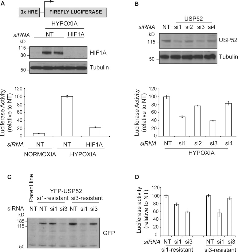
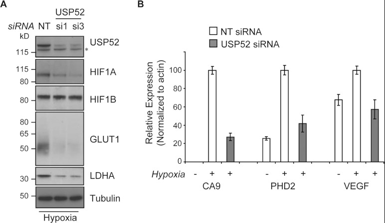
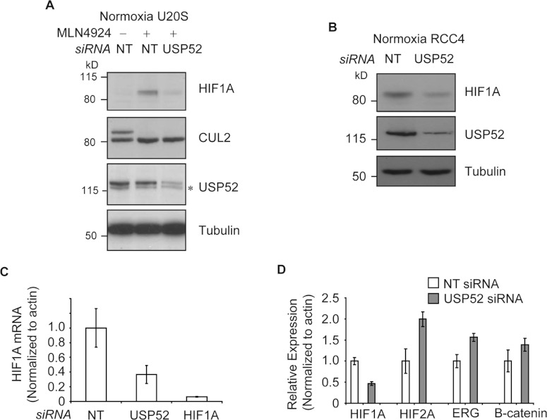
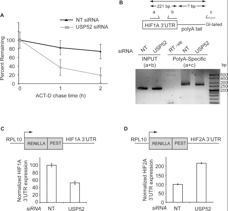
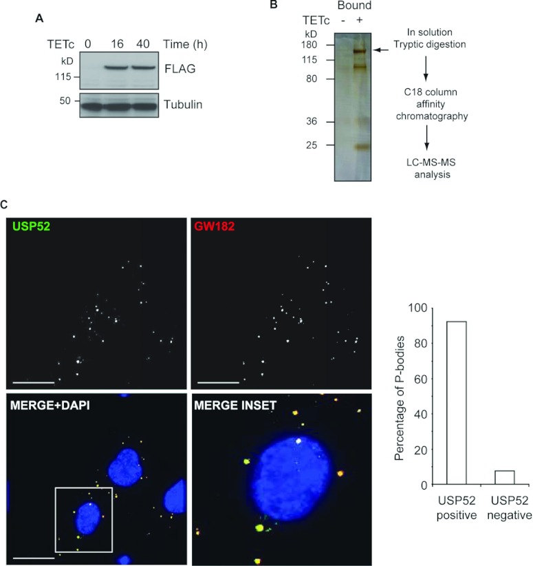
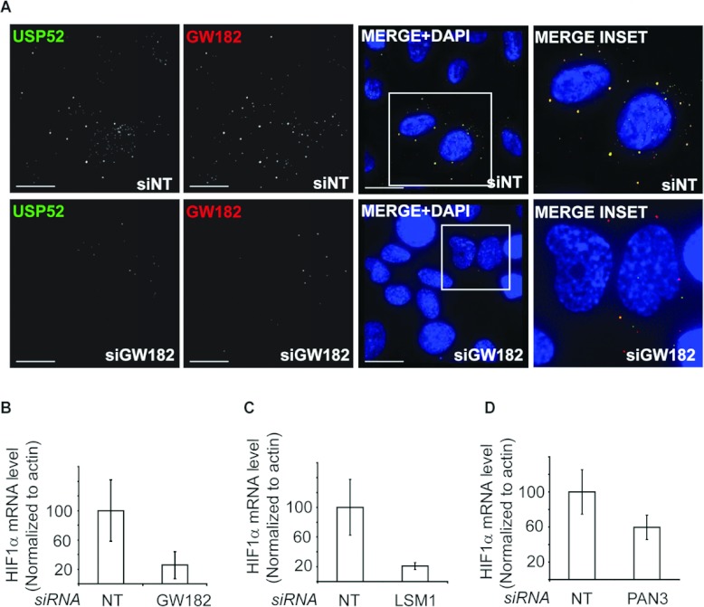
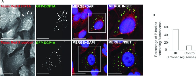
HIF1A (hypoxia-inducible factor 1α) is the master regulator of the cellular response to hypoxia and is implicated in cancer progression. Whereas the regulation of HIF1A protein in response to oxygen is well characterized, less is known about the fate of HIF1A mRNA. In the present study, we have identified the pseudo-DUB (deubiquitinating enzyme)/deadenylase USP52 (ubiquitin-specific protease 52)/PAN2 [poly(A) nuclease 2] as an important regulator of the HIF1A-mediated hypoxic response. Depletion of USP52 reduced HIF1A mRNA and protein levels and resulted in reduced expression of HIF1A-regulated hypoxic targets due to a 3'-UTR (untranslated region)-dependent poly(A)-tail-length-independent destabilization in HIF1A mRNA. MS analysis revealed an association of USP52 with several P-body (processing body) components and we confirmed further that USP52 protein and HIF1A mRNA co-localized with cytoplasmic P-bodies. Importantly, P-body dispersal by knockdown of GW182 or LSM1 resulted in a reduction of HIF1A mRNA levels. These data uncover a novel role for P-bodies in regulating HIF1A mRNA stability, and demonstrate that USP52 is a key component of P-bodies required to prevent HIF1A mRNA degradation.


Figures

Figure 1
Figure 1. USP52 is a regulator of the hypoxia response
(A) U2OS osteosarcoma cells stably expressing a hypoxia reporter construct consisting of three tandem HREs fused to the firefly luciferase gene (U2OS-HRE) were used to identify novel mediators of the hypoxia response. Western blot analysis demonstrates that HIF1A protein expression increases concomitant with a 10-fold increase in luciferase activity upon hypoxia treatment. Tubulin was used as a loading control. (B) Deconvolution analysis of USP52 revealed that individual siRNAs si1 and si3 both elicited an impaired response to hypoxia that correlated with the ability of these oligonucleotides to reduce USP52 protein levels by approximately 50%. Tubulin was used as a loading control. (C) Western blot of U2OS-HRE cells stably expressing siRNA-resistant YFP–USP52 were resistant to the individual siRNA as indicated, but remained sensitive to the other siRNA. (D) Luciferase assays of siRNA-resistant cells lines revealed USP52 si1 treatment was rescued to 80%, whereas si3 treatment was rescued to 95%. Cell lines remained sensitive to the individual siRNA to which resistance was not designed. Molecular masses are indicated in kDa in the blots, and results in histograms are means±S.E.M.
Figure 2
Figure 2. USP52 potentiates the hypoxic response
(A) Depletion of USP52 using siRNA si1 or si3 in U2OS cells reduces the levels of HIF1A, but not HIF1B, protein as assessed by Western blot analysis. HIF1A transcriptional targets GLUT1 and LDHA also showed decreased protein expression. Tubulin was used as a loading control. Molecular masses are indicated in kDa. (B) Real-time RT–PCR analysis shows that the expression of HIF1A targets CA9, PHD2 and VEGF are all induced in U2OS cells in response to hypoxia. USP52 knockdown reduces the ability of cells to increase expression of all HIF1A target genes. Levels were normalized to β-actin. Results are means±S.E.M.
Figure 3
Figure 3. USP52 regulates the hypoxia pathway in a VHL-independent manner by controlling HIF1A mRNA levels
(A) Normoxic U2OS cells were treated with MLN4924 to block CUL2 NEDD8ylation and inactivate the VHL complex. USP52 depletion was sufficient to decrease HIF1A protein levels, demonstrating independence of the VHL complex. Tubulin was used as a loading control. (B) USP52 depletion in VHL-deficient RCC4 renal cancer cells reduced HIF1A protein levels under normoxia. Tubulin was used as a loading control. (C) U2OS cells were treated with siRNA against USP52 and HIF1A, and the levels of HIF1A mRNA were assessed by real-time RT–PCR analysis. HIF1A siRNA reduced HIF1A mRNA levels to below 10%, whereas USP52 depletion reduced HIF1A mRNA to approximately 40%. Levels were normalized to β-actin. (D) USP52 was depleted in HeLa cells which were subject to real-time RT–PCR analysis. HIF1A mRNA levels were reduced by approximately 50% in HeLa cells by USP52 knockdown. HIF2A levels were increased 2-fold upon USP52 knockdown, whereas ERG and CTNNB1 mRNA levels were both increased 1.5-fold upon USP52 depletion. Levels were normalized to β-actin. Molecular masses are indicated in kDa in the blots, and results in histograms are means±S.E.M.
Figure 4
Figure 4. USP52 is required for stability of HIF1A mRNA
(A) Actinomycin D (ACT-D) chase experiments were performed in U2OS cells treated with either NT or USP52 siRNA. Real-time RT–PCR analysis revealed that depletion of USP52 decreased HIF1A mRNA half-life from 214 min to 35 min. Initial mRNA levels were normalized to 100% to account for lower HIF1A levels in USP52-depleted cells. Levels were normalized to β-actin. (B) U2OS cells were treated with NT or USP52 siRNA, and cDNA was made from G/I-tailed mRNA. PCR was performed using primers within HIF1A 3′-UTR (a+b) to generate a 221 bp product, and in a separate reaction with forward primer (a) and universal reverse primer (c). Poly(A) tail length in both NT and USP52 siRNA-treated cells was found to be predominantly ~60 residues long, calculated by subtracting the size of the (a+b) reaction product from the size of the (a+c) reaction product. Sizes are indicated in bp. (C) HEK-293 cells depleted of USP52 were transfected with the HIF1A 3′-UTR Renilla luciferase reporter shown. USP52 knockdown caused a 50% decrease in the expression of HIF1A 3′-UTR construct. Renilla luciferase values were normalized to firefly luciferase to control for transfection efficiency. (D) HEK-293 cells depleted of USP52 were transfected with the HIF2A 3′-UTR Renilla luciferase reporter shown. HIF2A 3′-UTR expression was increased 2-fold upon USP52 depletion. Renilla luciferase values were normalized to firefly luciferase to control for transfection efficiency. Results are means±S.E.M.
Figure 5
Figure 5. USP52 interacts with P-body components
(A) HEK-293 cells expressing tetracycline (TETc)-inducible FLAG–USP52 were generated. USP52 expression was induced by 16 or 40 h of TETc treatment as assessed by Western blot analysis. Tubulin was used as a loading control. (B) Inducible USP52 cells were either untreated or treated with TETc before FLAG–USP52 immunopreciptation. A fraction of eluted proteins (1/20th) were separated by denaturing gel electrophoresis and silver-stained. The position of FLAG–USP52 is indicated by an arrow. The remaining volume of eluted proteins was prepared for LC–MS/MS analysis as indicated in the flow chart. (C) Co-immunofluorescent staining of U2OS cells with USP52 and the P-body marker GW182 confirms that USP52 is a component of P-bodies. Quantification revealed that over 90% of P-bodies were USP52-positive, where 468 P-bodies were counted from 113 cells. Scale bars, 30 μm. Cells were counterstained with DAPI (4′,6-diamidino-2-phenylindole). Molecular masses are indicated in kDa in the blots.
Figure 6
Figure 6. GW182 maintains P-body integrity and HIF1A mRNA levels
(A) U2OS cells treated with either NT or GW182 siRNA were co-immunostained with USP52 and GW182. GW182 depletion by siRNA reduced the total number of P-bodies and dispersed USP52 from foci. Scale bars, 30 μm. Cells were counterstained with DAPI (4′,6-diamidino-2-phenylindole). (B) GW182 depletion caused a reduction in HIF1A mRNA levels to approximately 20%. Levels were normalized to β-actin. (C) LSM1 depletion caused a reduction in HIF1A mRNA levels. (D) PAN3 depletion caused a reduction in HIF1A mRNA levels. Levels were normalized to β-actin. Results in histograms are means±S.E.M.
Figure 7
Figure 7. HIF1A mRNA localizes to P-bodies
(A) U2OS cells were transfected with GFP–DCP1A, and HIF1A mRNA localization was assessed by FISH analysis with Texas-Red-X-labelled antisense probes. HIF1A was found to co-localize to GFP–DCP1A-positive P-bodies, whereas only background co-localization was observed with sense control probes. Scale bars, 30 μm. Nuclei were counterstained with DAPI (4′,6-diamidino-2-phenylindole). (B) The number of GFP–DCP1A-positive P-bodies containing HIF1A fluorescence (n=284) compared with sense control (n=489) were determined.

References

    1. Kenneth N. S., Rocha S. Regulation of gene expression by hypoxia. Biochem. J. 2008;414:19–29. - PubMed
    1. Salceda S., Caro J. Hypoxia-inducible factor 1α (HIF-1α) protein is rapidly degraded by the ubiquitin–proteasome system under normoxic conditions: its stabilization by hypoxia depends on redox-induced changes. J. Biol. Chem. 1997;272:22642–22647. - PubMed
    1. Keith B., Johnson R. S., Simon M. C. HIF1α and HIF2α: sibling rivalry in hypoxic tumour growth and progression. Nat. Rev. Cancer. 2012;12:9–22. - PMC - PubMed
    1. Maxwell P. H., Wiesener M. S., Chang G. W., Clifford S. C., Vaux E. C., Cockman M. E., Wykoff C. C., Pugh C. W., Maher E. R., Ratcliffe P. J. The tumour suppressor protein VHL targets hypoxia-inducible factors for oxygen-dependent proteolysis. Nature. 1999;399:271–275. - PubMed
    1. Epstein A. C., Gleadle J. M., McNeill L. A., Hewitson K. S., O’Rourke J., Mole D. R., Mukherji M., Metzen E., Wilson M. I., Dhanda A., et al. C. elegans EGL-9 and mammalian homologs define a family of dioxygenases that regulate HIF by prolyl hydroxylation. Cell. 2001;107:43–54. - PubMed

Publication types

MeSH terms