. 2013 Jul 1;22(13):1921-31.
doi: 10.1089/scd.2012.0575. Epub 2013 Apr 5.

Reconstitution of the myocardium in regenerating newt hearts is preceded by transient deposition of extracellular matrix components

Affiliations

Reconstitution of the myocardium in regenerating newt hearts is preceded by transient deposition of extracellular matrix components

Tanja Piatkowski et al. Stem Cells Dev. .

Abstract

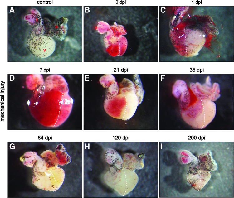
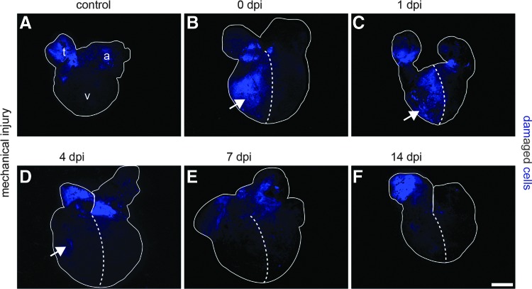
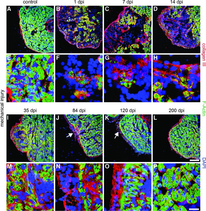
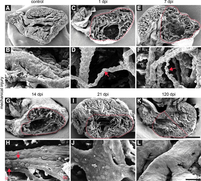
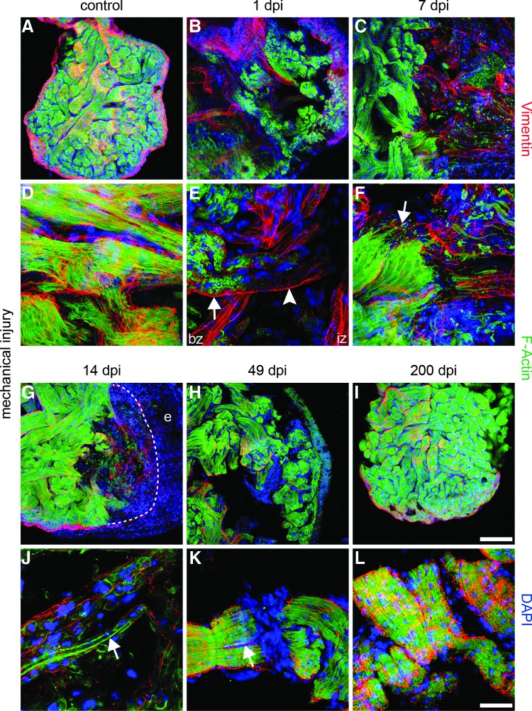
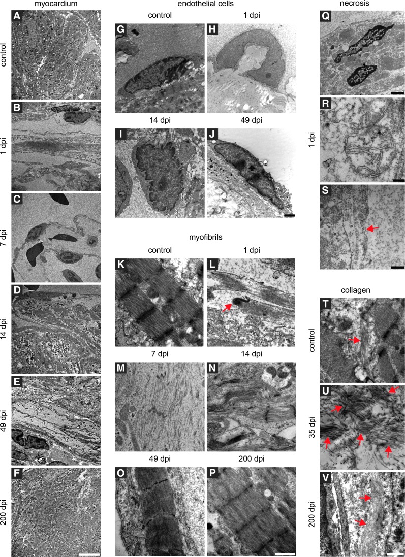
Adult newts efficiently regenerate the heart after injury in a process that involves proliferation of cardiac muscle and nonmuscle cells and repatterning of the myocardium. To analyze the processes that underlie heart regeneration in newts, we characterized the structural changes in the myocardium that allow regeneration after mechanical injury. We found that cardiomyocytes in the damaged ventricle mainly die by necrosis and are removed during the first week after injury, paving the way for the extension of thin myocardial trabeculae, which initially contain only very few cardiomyocytes. During the following 200 days, these thin trabeculae fill up with new cardiomyocytes until the myocardium is fully reconstituted. Interestingly, reconstruction of the newly formed trabeculated network is accompanied by transient deposition of extracellular matrix (ECM) components such as collagen III. We conclude that the ECM is a critical guidance cue for outgrowing and branching trabeculae to reconstruct the trabeculated network, which represents a hallmark of uninjured cardiac tissue in newts.


Figures

FIG. 1.
FIG. 1.
Macroscopic overview of injured and regenerating newt hearts. (A) The newt heart consists of an atrium (a), a ventricle (v), and an aortic trunk (t). The surface is partially covered by melanocytes. (B) Immediately after mechanical injury of the right half of the ventricle (0 dpi), the injured region appears red, probably due to restricted blood transport. (C–E) Restricted blood flow is visible up to 21 dpi when the size of the injured area decreases. (F) Further improvement of the injured area five weeks after damage. (G–I) At 84 dpi and afterward, no macroscopic differences between the damaged and undamaged hearts are visible. Dashed lines separate the injured from the uninjured region.
FIG. 2.
FIG. 2.
Mechanical injury of the newt ventricle causes damage of the plasma membranes. The cell membrane-impermeable dye SYTOX Blue stains DNA of cells with damaged membranes. (A) Control hearts show no staining of cells in the ventricle (v), but signals in the atrium (a) and the aortic trunk (t), which is caused by preparation artifacts. (B) The right half of the ventricle contains numerous SYTOX Blue-labeled cells immediately after injury and one day later (C), indicating a high degree of damage. (D) Three days later, only a few SYTOX Blue-labeled cells remain. (E–F) After 7 dpi, no SYTOX Blue-labeled cells are present in the injured area anymore. Dashed lines separate the injured from the uninjured region; Arrows indicate SYTOX Blue-positive cells in the ventricle. Scale bar: 600 μm.
FIG. 3.
FIG. 3.
Increased deposition of extracellular matrix (ECM) during newt heart regeneration. (A, E) Control hearts display collagen III immunofluorescence around F-actin-positive cardiomyocytes and underneath the epicardium. Additionally, collagen III covers the ventricular lumen. (B, F) Increase of collagen III deposition at the outside of the injured region and around remaining cardiomyocytes at 1 dpi. (C, D, G, H, I, M) Collagen III deposits increase up to 35 dpi. Increased signal intensity is obvious around small F-actin-positive patches. (J, N) Reduced presence of collagen III after 84 dpi mainly in the outer myocardium. (K, O) After 120 dpi, collagen III levels decline with the exception of some deposits at the epicardium and the subjacent myocardium. (L, P) At 200 dpi, no significant differences to the control hearts are visible anymore. Arrows indicate the regions with higher collagen III levels at 84 and 120 dpi. F-actin: green; collagen III: red; DAPI: blue; scale bar: (A–D; I–L) 300 μm; (E–H; M–P) 50 μm.
FIG. 4.
FIG. 4.
Large structural defects in the myocardium after mechanical injury. (A, B) Scanning electron microscopy analysis of newt hearts reveals a small ventricular lumen and a compact trabeculated myocardium with a smooth surface. Individual trabeculae cross the ventricle. (C, D) At 1 dpi, a partial loss of cardiac tissue and a decrease in size of trabeculae in the injured region are apparent. (E, F) Large structural defects are visible at 7 dpi. The size of the remaining trabeculae is reduced, exposing the nuclei of subjacent cells (red arrows indicate thin trabeculae after 1 and 7 dpi). (G, H) At 14 dpi, tissue defects are still apparent. Parts of trabeculae in the border zone (bz) have regained a normal morphology, while the surface of trabeculae in the injured zone (iz) still show off the nuclei of underlying cells (red arrows indicate the cells under the surface of trabeculae in the injured region) (I, J) At 21 dpi, subjacent cells are still visible at the surface of the trabeculae, but the overall trabecular surface has smoothened. The injured region is replenished with cardiac tissue, but still shows gaps. (K, L) At 120 dpi, the myocardium shows a compact organization. No significant differences compared to the control hearts are evident. Red dashed lines indicate the injured region; scale bar: (A, C, E, G, I, K) 500 μm; (B, D, F, H, J, L) 50 μm.
FIG. 5.
FIG. 5.
Repopulation of remaining trabeculae by immature cardiomyocytes. (A, D) Immunofluorescence staining of the newt myocardium reveals a compact network of trabeculae filled with F-actin-positive cardiomyocytes and parallel-organized myofilaments. Cardiomyocytes are covered by Vimentin-positive endothelial cells. (B, E) At 1 dpi, the injured area contains disorganized trabeculae. Cardiomyocytes in the border zone (bz) shows an irregular pattern of F-actin staining (arrow) with partial loss of the F-actin signal inside the remaining trabeculae of the injured zone (iz) (arrow head). (C, F) Decline of F-actin expression in the iz at 7 dpi. Small F-actin-positive processes project from the border zone into the injured area (arrow). (G, J) At 14 dpi, the remaining trabeculae fill with F-actin-positive cells containing rare, thin myofibrils (arrow). (H, K) After 49 dpi, F-actin-positive myofibrils repattern, but gaps in the myocardium are still apparent. Some trabeculae split into two thinner branches, which are both covered by Vimentin-positive cells (arrow). (I, L) At 200 dpi, regeneration is completed, and the myocardium has regained a compact network of trabeculae with well-organized cardiomyocytes covered by Vimentin-positive cells. F-actin: green; Vimentin: red; DAPI: blue; e: epicardium. Scale bar A–C & G–I: 300 μm; D–F & J–L: 50 μm.
FIG. 6.
FIG. 6.
Ultrastructural changes of the newt myocardium after mechanical damage. (A-F) Transmission electron microscopy analysis of the regenerating myocardium. (A) The undamaged myocardium of the ventricle shows compact organization of cardiomyocytes inside the trabeculae. (B) At 1 dpi, reduction of cardiomyocytes inside trabeculae is apparent. The majority of damaged cardiomyocytes is removed until 7 dpi (C). (D) At 14 dpi, repatterning of ventricular structures has started as indicated by repopulation of the remaining trabeculae with cardiomyocytes. Gaps in the myocardium and inside trabeculae are still visible. At 49 dpi, regeneration led to continuous improvement of tissue morphology. Most trabeculae are populated by cardiomyocytes with only few exceptions (E). (F) At 200 dpi, regeneration is completed. No differences between the injured and uninjured hearts are visible. (G-J) Morphological alterations of endothelial cells immediately after damage. (G) Endothelial cells in the control hearts are flat with elliptic nuclei. (H) At 1 dpi, the nuclei of endothelial cells in the injured area are arched and spherical. After 14 dpi, the nuclei of endothelial cells regain a normal round (I) to elliptic shape, which is maintained at later stages (J). (K-P) Mechanical injury induces massive damage of myofibrils inside cardiomyocytes. (K) Myofibrils of the hearts without injury display clearly defined sarcomeres as indicated by the typical pattern of Z-lines, I and M bands, as well as H & A zones. (L) Loss of sarcomere integrity and intercalated disks 1 dpi (red arrow). (M) Fusiform myofibrils without intact Z-lines after 7 dpi. (N) Some myofibrils in the border zone contain no Z-lines, although myofibrils are aligned to myofibrils with Z-lines. (O) At 49 dpi, myofibrils show a normal morphology despite some gaps in the myocardium. (P) Regeneration is complete after 200 dpi. (Q-S) Necrosis of different cell types after mechanical injury of the ventricle. (Q) At 1 dpi, cardiomyocytes in the injured area contain necrotic nuclei and (R) swollen mitochondria with fewer and smaller cristae. (S) Membrane integrity of cardiomyocytes and endothelial cells is compromised (indicated by red arrows). (T–V) Changes in collagen deposition during regeneration. (T) The control hearts maintain low levels of collagen fibres in the interstitial space. (U) Increasing deposition of collagen fibres until 35 dpi. After 35 dpi, the collagen level drops until normal concentrations are reached at 200 dpi when regeneration is complete (collagen fibres are indicated by red arrows) (V). Scale bars: A–F: 10 μm; G–H: 2 μm; K–P: 1 μm; Q: 2 μm; R: 200 nm; S: 500 nm; T–V: 1 μm

Similar articles

Cited by

References

    1. Jourdan-Lesaux C. Zhang J. Lindsey ML. Extracellular matrix roles during cardiac repair. Life Sci. 2010;87:391–400. - PMC - PubMed
    1. Zamilpa R. Lindsey ML. Extracellular matrix turnover and signaling during cardiac remodeling following MI: causes and consequences. J Mol Cell Cardiol. 2010;48:558–563. - PMC - PubMed
    1. Nunes SS. Song H. Chiang CK. Radisic M. Stem cell-based cardiac tissue engineering. J Cardiovasc Transl Res. 2011;4:592–602. - PubMed
    1. Baddour JA. Sousounis K. Tsonis PA. Organ repair and regeneration: an overview. Birth Defects Res C Embryo Today. 2012;96:1–29. - PubMed
    1. Laflamme MA. Murry CE. Heart regeneration. Nature. 2011;473:326–335. - PMC - PubMed

Publication types

Substances

LinkOut - more resources