

TIMP-2 mutant decreases MMP-2 activity and augments pressure overload induced LV dysfunction and heart failure
- PMID: 23398532
- PMCID: PMC3881363
- DOI: 10.3109/13813455.2012.755548
TIMP-2 mutant decreases MMP-2 activity and augments pressure overload induced LV dysfunction and heart failure
Abstract
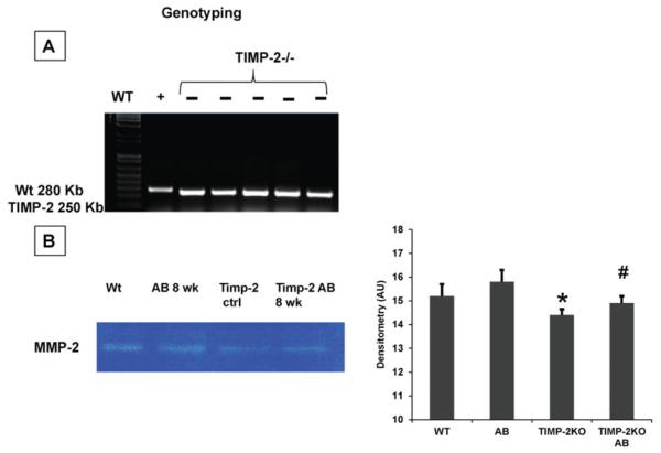
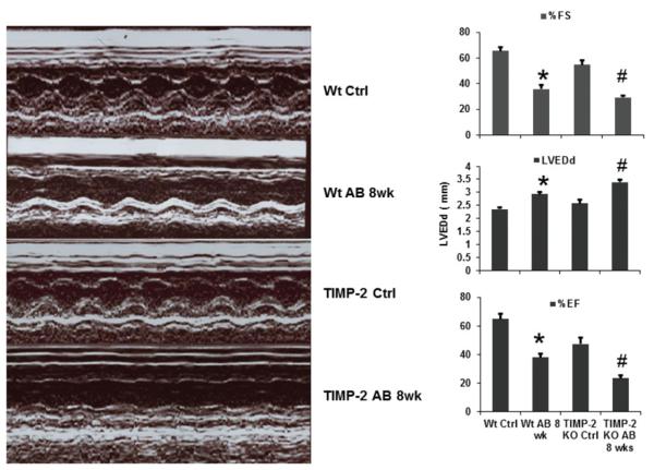
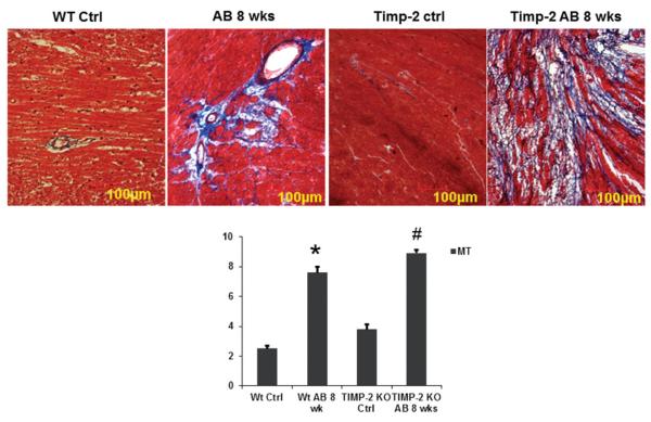
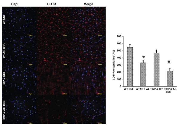
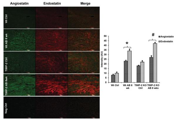
Pressure overload induces cardiac extracellular matrix (ECM) remodelling and results in heart failure. ECM remodelling by matrix metalloproteinases (MMPs) is primarily regulated by their target inhibitors, tissue inhibitor of matrix metalloproteinases (TIMPs). It is known that TIMP-2 is highly expressed in myocardium and is required for cell surface activation of pro-MMP-2. We and others have reported that imbalance between angiogenic growth factors and anti-angiogenic factors results in transition from compensatory cardiac hypertrophy to heart failure. We previously reported the pro-angiogenic role of MMP-2 in cardiac compensation, however, the specific role of TIMP-2 during pressure overload is yet unclear. We hypothesize that genetic ablation of TIMP-2 exacerbates the adverse cardiac matrix remodelling due to lack of pro-angiogenic MMP-2 and increase in anti-angiogenic factors during pressure overload stress and results in severe heart failure. To verify this, ascending aortic banding (AB) was created to mimic pressure overload, in wild type C57BL6/J and TIMP-2-/- (model of MMP-2 deficiency) mice. Left ventricular (LV) function assessed by echocardiography and pressure-volume loop studies showed severe LV dysfunction in TIMP-2-/- AB mice compared to controls. Expression of MMP-2, vascular endothelial growth factor (VEGF) was decreased and expression of MMP-9, anti-angiogenic factors endostatin and angiostatin was increased in TIMP-2-/- AB mice compared with wild type AB mice. Connexins (Cx) are the gap junction proteins that are widely present in the myocardium and play an important role in endothelial-myocyte coupling. Our results showed that expression of Cx 37 and 43 was decreased in TIMP-2-/- AB mice compared with corresponding wild type controls. These results suggest that genetic ablation of TIMP-2 decrease the expression of pro-angiogenic MMP-2, VEGF and increases anti-angiogenic factors that results in exacerbated abnormal ventricular remodelling leading to severe heart failure.
Figures


Similar articles
-
MMP-2/TIMP-2/TIMP-4 versus MMP-9/TIMP-3 in transition from compensatory hypertrophy and angiogenesis to decompensatory heart failure.Arch Physiol Biochem. 2010 May;116(2):63-72. doi: 10.3109/13813451003652997. Arch Physiol Biochem. 2010. PMID: 20230216 Free PMC article.
-
Mitochondrial division/mitophagy inhibitor (Mdivi) ameliorates pressure overload induced heart failure.PLoS One. 2012;7(3):e32388. doi: 10.1371/journal.pone.0032388. Epub 2012 Mar 27. PLoS One. 2012. PMID: 22479323 Free PMC article.
-
Combination of tumor necrosis factor-alpha ablation and matrix metalloproteinase inhibition prevents heart failure after pressure overload in tissue inhibitor of metalloproteinase-3 knock-out mice.Circ Res. 2005 Aug 19;97(4):380-90. doi: 10.1161/01.RES.0000178789.16929.cf. Epub 2005 Jul 21. Circ Res. 2005. PMID: 16037568
-
Matrix Metalloproteinases and Tissue Inhibitors of Metalloproteinases in Extracellular Matrix Remodeling during Left Ventricular Diastolic Dysfunction and Heart Failure with Preserved Ejection Fraction: A Systematic Review and Meta-Analysis.Int J Mol Sci. 2020 Sep 14;21(18):6742. doi: 10.3390/ijms21186742. Int J Mol Sci. 2020. PMID: 32937927 Free PMC article.
-
Extracellular matrix remodeling during the progression of volume overload-induced heart failure.J Mol Cell Cardiol. 2010 Mar;48(3):564-9. doi: 10.1016/j.yjmcc.2009.06.001. Epub 2009 Jun 11. J Mol Cell Cardiol. 2010. PMID: 19524591 Free PMC article. Review.
Cited by
-
The Role of Connexin-43 in the Inflammatory Process: A New Potential Therapy to Influence Keratitis.J Ophthalmol. 2019 Jan 21;2019:9312827. doi: 10.1155/2019/9312827. eCollection 2019. J Ophthalmol. 2019. PMID: 30805212 Free PMC article. Review.
-
Resuscitation of a dead cardiomyocyte.Heart Fail Rev. 2015 Nov;20(6):709-19. doi: 10.1007/s10741-015-9501-z. Heart Fail Rev. 2015. PMID: 26311463 Review.
-
Homocysteine as a Predictor of Paroxysmal Atrial Fibrillation-Related Events: A Scoping Review of the Literature.Diagnostics (Basel). 2022 Sep 9;12(9):2192. doi: 10.3390/diagnostics12092192. Diagnostics (Basel). 2022. PMID: 36140593 Free PMC article.
-
Cellular mechanisms and molecular pathways linking bitter taste receptor signalling to cardiac inflammation, oxidative stress, arrhythmia and contractile dysfunction in heart diseases.Inflammopharmacology. 2023 Feb;31(1):89-117. doi: 10.1007/s10787-022-01086-9. Epub 2022 Dec 6. Inflammopharmacology. 2023. PMID: 36471190 Free PMC article. Review.
-
Regulation and involvement of matrix metalloproteinases in vascular diseases.Front Biosci (Landmark Ed). 2016 Jan 1;21(1):89-118. doi: 10.2741/4378. Front Biosci (Landmark Ed). 2016. PMID: 26709763 Free PMC article. Review.
References
-
- Alexander SM, Jackson KJ, Bushnell KM, McGuire PG. Spatial and temporal expression of the 72-kDa type IV collagenase (MMP-2) correlates with development and differentiation of valves in the embryonic avian heart. Dev Dyn. 1997;209:261–8. - PubMed
-
- Braunwald E. Heart disease: A textbook of cardiovascular medicine. W.B. Saunders; Philadelphia, PA: 1980.
-
- Butler GS, Butler MJ, Atkinson SJ, et al. The TIMP2 membrane type 1 metalloproteinase “receptor” regulates the concentration and efficient activation of progelatinase A. A kinetic study. J Biol Chem. 1998;273:871–80. - PubMed
Publication types
MeSH terms
Substances
Grants and funding
LinkOut - more resources
Full Text Sources
Other Literature Sources
Medical
Research Materials
Miscellaneous