. 2013 Feb 12;11(1):4.
doi: 10.1186/1546-0096-11-4.

Coronary artery involvement in pediatric Takayasu's arteritis: Case report and literature review

Affiliations

Coronary artery involvement in pediatric Takayasu's arteritis: Case report and literature review

Shaun Mohan et al. Pediatr Rheumatol Online J. .

Abstract

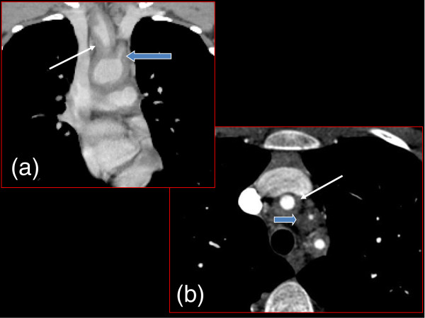
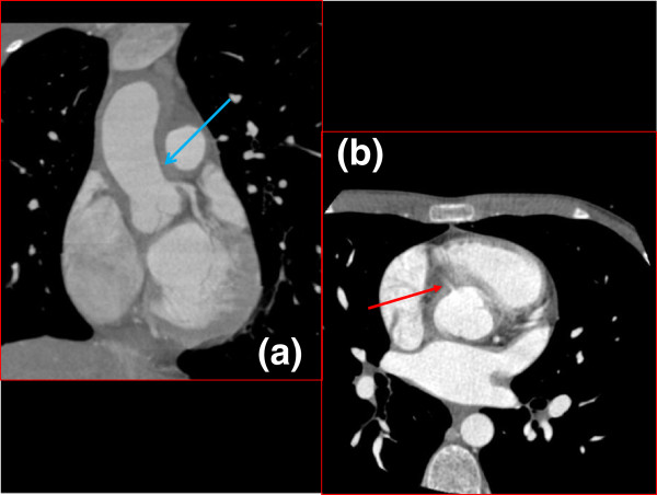
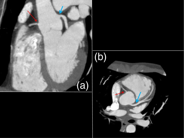
Takayasu's arteritis (TA) is a form of chronic vasculitis that typically occurs in young adult Asian females, but it can also present in younger patients not fitting this classic profile. In these cases, the sequelae are generally similar to those found in adults. The disease predominantly affects the aorta and its primary branches. However, the coronary arteries are also affected in up to 20% of cases, which may precipitate myocardial infarction. Imaging of the coronary arteries therefore becomes critically important in the evaluation of a patient with possible Takayasu's arteritis. We present a case of a pediatric patient with TA who had no symptoms of angina but who was found to have significant coronary artery involvement on diagnostic imaging. This necessitated tailoring of traditional management.


Figures

Figure 1
Figure 1
Coronal (a) and Axial (b) CT Chest. Concentric wall thickening at the origin of the major aortic arch branches: brachio-cephalic trunk (thin white arrow) and left common carotid artery origin (thick blue arrow).
Figure 2
Figure 2
Coronal (a) and Axial (b) Cardiac Gated CT Chest. Fifty percent narrowing of the origin of the left coronary artery is demonstrated by arrow in (a, blue arrow). The right coronary artery should also be visible in this coronal section but was not due to stenosis (a). It can be seen as a wisp coming off the ascending aorta in the axial image (b, red arrow).
Figure 3
Figure 3
Coronal (a) and Axial (b) Cardiac Gated CT Chest 4 months after systemic therapy. The right coronary artery, though still stenotic, was improved and now able to be visualized on coronal section (a, thin red arrow). The left coronary artery opened up significantly (b, blue arrow).

References

    1. Miller M, Pachman L. In: Nelson Textbook of Pediatrics. 18. Kleigman R, Jenson H, Behrman R, Stanton B, editor. Philadelphia: Saunders; 2007. Vasculitis Syndromes; pp. 1045–1046.
    1. Brunner J, Feldman BM, Tyrrell PN, Kuemmerle-Deschner JB, Zimmerhackl LB, Gassner I, Benseler SM. Takayasu arteritis in children and adolescents. Rheumatology (Oxford) 2010;49(10):1806–1814. doi: 10.1093/rheumatology/keq167. - DOI - PubMed
    1. Tann OR, Tulloh RM, Hamilton MC. Takayasu’s disease: a review. Cardiol Young. 2008;18(3):250–259. - PubMed
    1. Arnaud L, Haroche J, Mathian A, Gorochov G, Amoura Z. Pathogenesis of Takayasu’s arteritis: a 2011 update. Autoimmun Rev. 2011;11(1):61–67. doi: 10.1016/j.autrev.2011.08.001. - DOI - PubMed
    1. Johnston SL, Lock RJ, Gompels MM. Takayasu arteritis: a review. J Clin Pathol. 2002;55(7):481–486. doi: 10.1136/jcp.55.7.481. - DOI - PMC - PubMed

LinkOut - more resources