

Attenuation of spinal cord injury-induced astroglial and microglial activation by repetitive transcranial magnetic stimulation in rats
- PMID: 23399872
- PMCID: PMC3565143
- DOI: 10.3346/jkms.2013.28.2.295
Attenuation of spinal cord injury-induced astroglial and microglial activation by repetitive transcranial magnetic stimulation in rats
Abstract
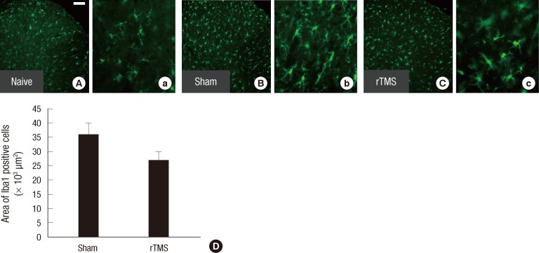
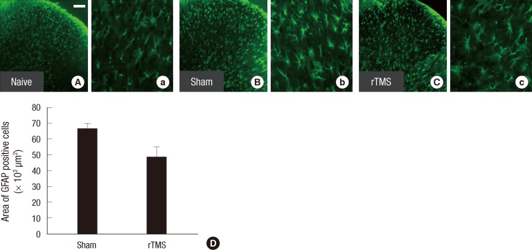
Spinal cord injury (SCI) causes not only loss of sensory and motor function below the level of injury but also chronic pain, which is difficult and challenging of the treatment. Repetitive transcranial magnetic stimulation (rTMS) to the motor cortex, of non-invasive therapeutic methods, has the motor and sensory consequences and modulates pain in SCI-patients. In the present study, we studied the effectiveness of rTMS and the relationship between the modulation of pain and the changes of neuroglial expression in the spinal cord using a rat SCI-induced pain model. Elevated expressions of Iba1 and GFAP, specific microglial and astrocyte markers, was respectively observed in dorsal and ventral horns at the L4 and L5 levels in SCI rats. But in SCI rats treated with 25 Hz rTMS for 8 weeks, these expressions were significantly reduced by about 30%. Our finding suggests that this attenuation of activation by rTMS is related to pain modulation after SCI. Therefore, rTMS might provide an alternative means of attenuating neuropathic pain below the level of SCI.
Keywords: Astrocytes; Microglia; Repetitive Transcranial Magnetic Stimulation; Spinal Cord Injury.
Figures
References
-
- Hulsebosch CE. From discovery to clinical trials: treatment strategies for central neuropathic pain after spinal cord injury. Curr Pharm Des. 2005;11:1411–1420. - PubMed
-
- Willis A, Mihalevich M, Neff RA, Mendelowitz D. Three types of postsynaptic glutamatergic receptors are activated in DMNX neurons upon stimulation of NTS. Am J Physiol. 1996;271:R1614–R1619. - PubMed
-
- Salter MW. Cellular neuroplasticity mechanisms mediating pain persistence. J Orofac Pain. 2004;18:318–324. - PubMed
-
- Wasner G, Lee BB, Engel S, McLachlan E. Residual spinothalamic tract pathways predict development of central pain after spinal cord injury. Brain. 2008;131:2387–2400. - PubMed
Publication types
MeSH terms
Substances
LinkOut - more resources
Full Text Sources
Other Literature Sources
Medical
Miscellaneous
