. 2013 Aug;27(8):1666-76.
doi: 10.1038/leu.2013.40. Epub 2013 Feb 12.

Gads (Grb2-related adaptor downstream of Shc) is required for BCR-ABL-mediated lymphoid leukemia

Affiliations

Gads (Grb2-related adaptor downstream of Shc) is required for BCR-ABL-mediated lymphoid leukemia

L C Gillis et al. Leukemia. 2013 Aug.

Abstract

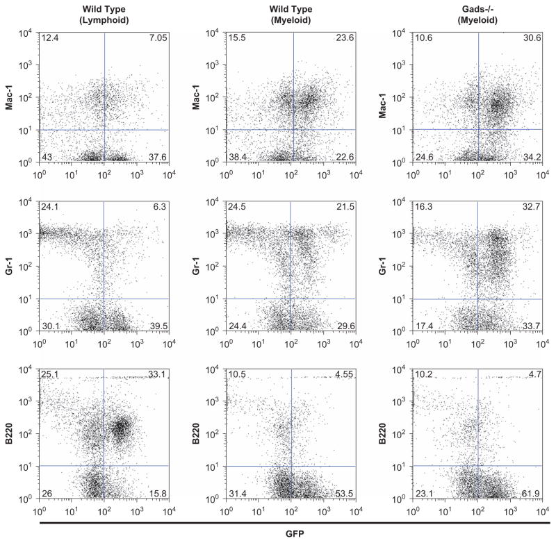
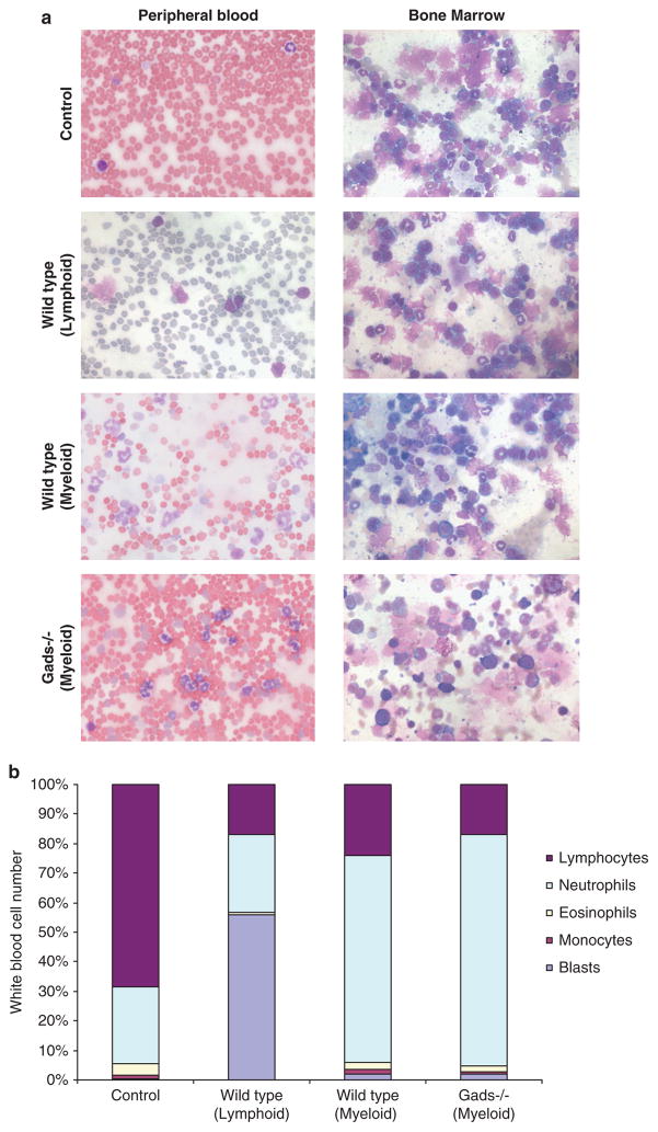
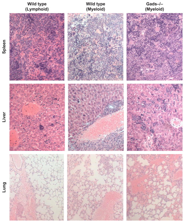
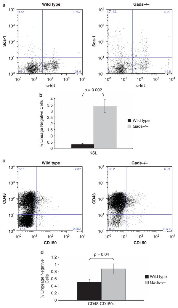
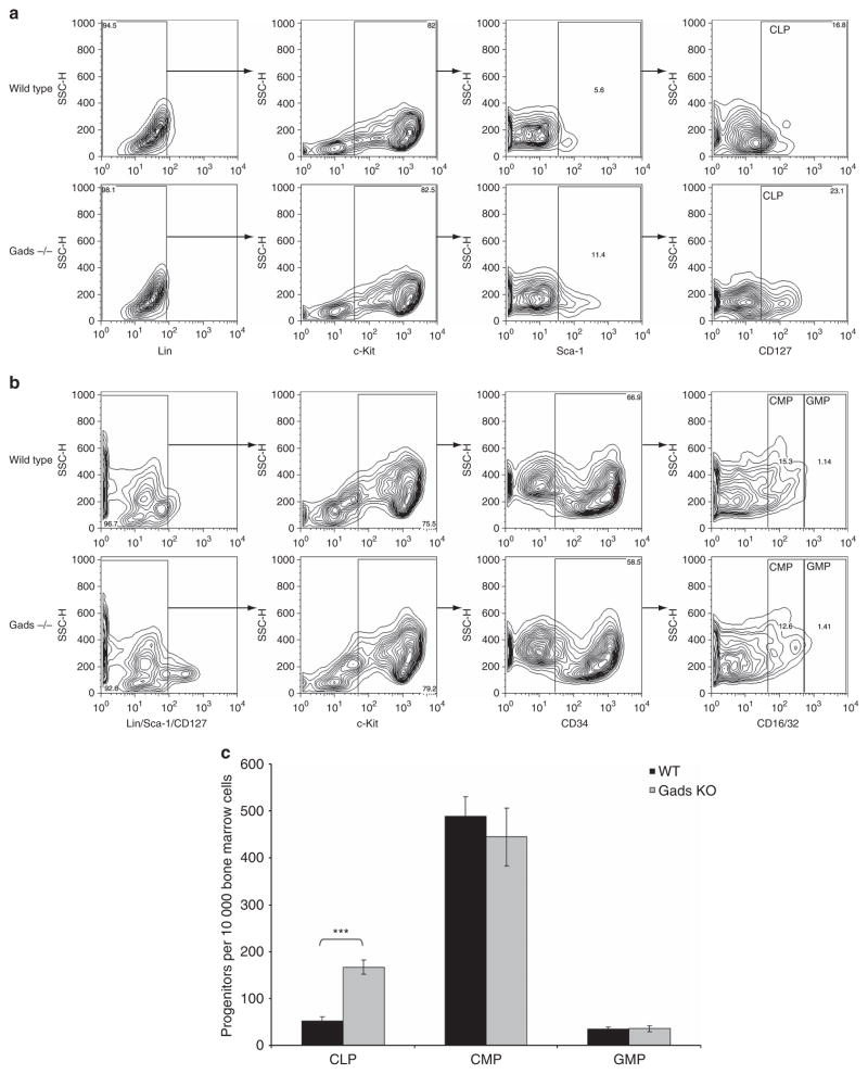
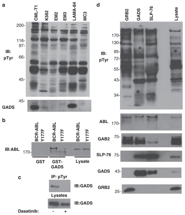
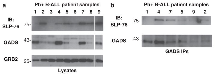
Philadelphia chromosome-positive leukemias, including chronic myeloid leukemia and B-cell acute lymphoblastic leukemia (B-ALL), are driven by the oncogenic BCR-ABL fusion protein. Animal modeling experiments utilizing retroviral transduction and subsequent bone marrow transplantation have demonstrated that BCR-ABL generates both myeloid and lymphoid disease in mice receiving whole bone marrow transduced with BCR-ABL. Y177 of BCR-ABL is critical to the development of myeloid disease, and phosphorylation of Y177 has been shown to induce GRB2 binding to BCR-ABL, followed by activation of the Ras and phosphoinositide 3 kinase signaling pathways. We show that the GRB2-related adapter protein, GADS, also associates with BCR-ABL, specifically through Y177 and demonstrate that BCR-ABL-driven lymphoid disease requires Gads. BCR-ABL transduction of Gads(-/-) bone marrow results in short latency myeloid disease within 3-4 weeks of transplant, while wild-type mice succumb to both a longer latency lymphoid and myeloid diseases. We report that GADS mediates a unique BCR-ABL complex with SLP-76 in BCR-ABL-positive cell lines and B-ALL patient samples. These data suggest that GADS mediates lymphoid disease downstream of BCR-ABL through the recruitment of specific signaling intermediates.


Conflict of interest statement

CONFLICT OF INTEREST

The authors declare no conflict of interest.

Figures

Figure 1
Figure 1
Gads is required for lymphoid disease induced by BCR-ABL. BCR-ABL-transduced bone marrow from non-5-FU donors was transplanted into wild-type recipient mice. Recipients of Gads −/− marrow showed reduced survival in comparison to recipients of WT marrow. The difference in survival is significant (P = 0.004), as determined by log-rank analysis.
Figure 2
Figure 2
BCR-ABL induces a mixed disease phenotype in recipients of non-5-FU-treated donor cells. Spleen cells from transplant recipients were analyzed by flow cytometry to determine the forward and side scatter profiles and the proportions of GFP-expressing B220 +, CD19 +, Gr-1 + and Mac-1 + cells. Wild-type recipients were diagnosed as either lymphoid (B220 + and CD19 +) or myeloid (Gr-1 + and Mac-1 +) whereas Gads-deficient recipients were diagnosed exclusively as myeloid.
Figure 3
Figure 3
Disease phenotypes are confirmed via histopathology and differential counting. (a) Histopathological analysis of peripheral blood and bone marrow was visualized by microscopic evaluation of slides stained with May-Grünwald and Giemsa. Original magnification × 400. (b) Differential counts were recorded for multiple animals (wild type (n = 5); Gads(−/−) (n = 8); for control non-transplanted mice (n = 4)) through counts of at least 200 total white blood cells in randomly selected sections of the peripheral blood smear. Average count of each compartment is reported.
Figure 4
Figure 4
Extramedullary hematopoiesis is found in bone marrow transplant recipients via histopathology. Spleen, liver and lung sections from transplant recipients were stained with hematoxylin and eosin and visualized by light microscopy. Original magnification (×200).
Figure 5
Figure 5
Stem cell progenitors are increased in Gads-deficient bone marrow. (a) Bone marrow cells were collected from 8-week-old wild-type (n = 6) and Gads-deficient mice (n = 6). Red blood cells were lysed and the remaining white blood cells were lineage depleted and incubated with fluorescent antibodies specific for cell surface markers kit (K), Sca-1 (S) and Lineage negative (Lin). Flow cytometry was used to enumerate specific cell types, representative samples are shown. (b) Data are presented as the average percentage of positive (lineage depleted) cells. Error bars represent standard error and significant differences, as measured by T test are noted. (c) Bone marrow cells were collected from 8-week-old, sex- and age-matched wild-type (n = 9) and Gads-deficient (n = 12) mice. Red blood cells were lysed and the remaining white blood cells were lineage depleted, then incubated with fluorescent antibodies specific for cell surface markers (CD48 and CD150). Flow cytometry was used to enumerate specific cell types. (d) Data are presented as the average percentage of positive (lineage depleted) cells. Error bars represent standard error and significant differences, as measured by T test, are indicated.
Figure 6
Figure 6
Increased CLP are observed in Gads-deficient bone marrow. Following lineage depletion, cells were incubated with fluorescent antibodies specific for cell surface markers expressed on CLP, CMP and GMP. Flow cytometry was used to enumerate specific cell types. The gating strategy utilized to identify (a) CLP, (b) CMP and (b) GMP is illustrated for representative wild-type and Gads −/− bone marrow. (c) Data are presented as the average number of CLP, CMP or GMP cells per 10 000 lineage-depleted bone marrow cells for wild-type (n = 12) and Gads-deficient mice (n = 12). Error bars represent standard error and significant differences, as measured by T test, are noted. (***P<0.0001).
Figure 7
Figure 7
GADS-SLP-76 are recruited to BCR-ABL Y177. (a) Several human leukemia cell lines were lysed in SDS-PAGE sample buffer and analyzed for levels of tyrosine-phosphorylated proteins via immunoblotting (IB). The blot was stripped and reprobed for GADS expression. (b) Pull-down experiments, with GST-Gads or GST alone, were performed using lysates from BaF/3 cells expressing wild-type BCR-ABL (wild type) or BCR-ABL Y177F (Y177F). The membrane was probed with a peptide-specific Abl antibody. (c) CML-T1 cells were incubated in RPMI or RPMI +Dasatinib. Tyrosine phosphorylation of GADS protein was detected by immunoprecipitations performed with the 4G10 anti-phosphotyrosine monoclonal antibody followed by western blot with anti-Gads antibody (upper panel). Lysates were probed with the anti-Gads antibody (lower panel). (d) CML-T1 cells were incubated with RMPI media (control), RPMI containing Dasatinib or RPMI containing R406. Lysates were immunoprecipitated with a GADS antibody and western blotting was completed with an anti-phosphotyrosine antibody and reprobed with a peptide-specific GADS antibody. GRB2, GADS and SLP-76 immunoprecipitations were performed on CML-T1 cell lysates and blotted with anti-phosphotyrosine. Proteins were detected by blotting with antibodies raised against ABL, GAB2, SLP-76, GADS and GRB2.
Figure 8
Figure 8
Gads is expressed in a complex with SLP-76 in a subset of Philadelphia chromosome-positive patient samples. (a) Lysates from Philadelphia chromosome-positive B-ALL patient samples were analyzed by western blot for expression of SLP-76, GADS and GRB2. (b) Anti-GADS immunoprecipitations (IPs) were performed on the B-ALL sample lysates that expressed GADS protein. The presence of SLP-76 protein was detected by western blot analysis.

Similar articles

Cited by

References

    1. Nowell PC, Hungerford DA. Chromosome studies on normal and leukemic human leukocytes. J Natl Cancer Inst. 1960;25:85–109. - PubMed
    1. Rowley JD. Chromosomal patterns in myelocytic leukemia. N Engl J Med. 1973;289:220–221. - PubMed
    1. Sawyers CL. Chronic myeloid leukemia. N Engl J Med. 1999;340:1330–1340. - PubMed
    1. O’Hare T, Zabriskie MS, Eiring AM, Deininger MW. Pushing the limits of targeted therapy in chronic myeloid leukaemia. Nat Rev Cancer. 2012;12:513–526. - PubMed
    1. Konopka JB, Watanabe SM, Witte ON. An alteration of the human c-abl protein in K562 leukemia cells unmasks associated tyrosine kinase activity. Cell. 1984;37:1035–1042. - PubMed

Publication types

MeSH terms

Substances