Meta-Analysis
. 2013 Nov;18(11):1225-34.
doi: 10.1038/mp.2013.1. Epub 2013 Feb 12.

Increased expression of BIN1 mediates Alzheimer genetic risk by modulating tau pathology

Affiliations
Meta-Analysis

Increased expression of BIN1 mediates Alzheimer genetic risk by modulating tau pathology

J Chapuis et al. Mol Psychiatry. 2013 Nov.

Abstract

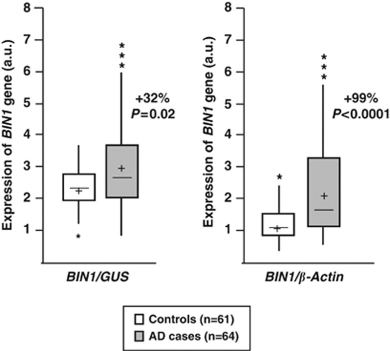
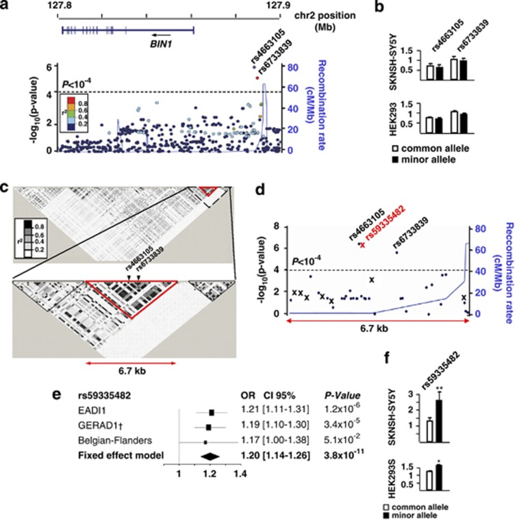
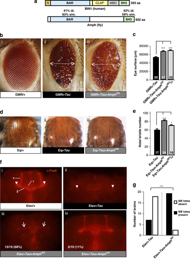
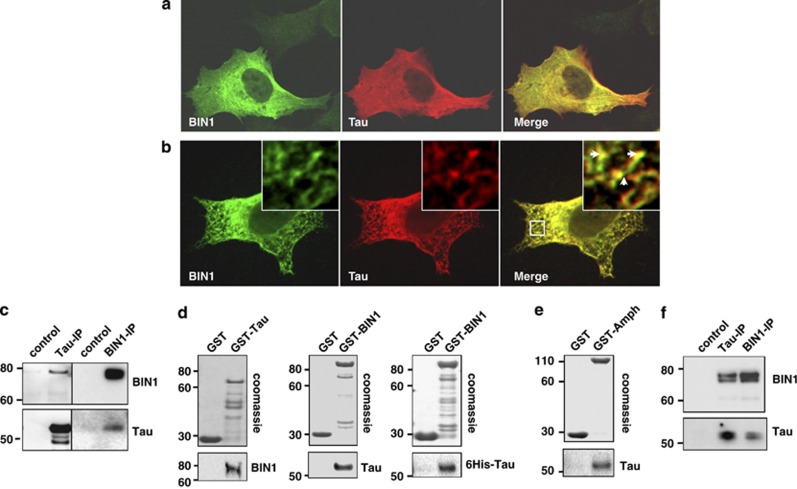
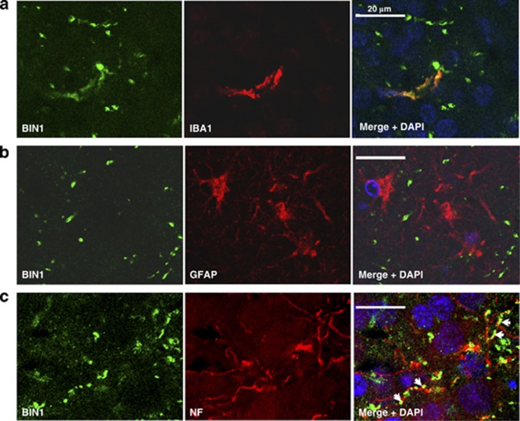
Genome-wide association studies (GWAS) have identified a region upstream the BIN1 gene as the most important genetic susceptibility locus in Alzheimer's disease (AD) after APOE. We report that BIN1 transcript levels were increased in AD brains and identified a novel 3 bp insertion allele ∼28 kb upstream of BIN1, which increased (i) transcriptional activity in vitro, (ii) BIN1 expression levels in human brain and (iii) AD risk in three independent case-control cohorts (Meta-analysed Odds ratio of 1.20 (1.14-1.26) (P=3.8 × 10(-11))). Interestingly, decreased expression of the Drosophila BIN1 ortholog Amph suppressed Tau-mediated neurotoxicity in three different assays. Accordingly, Tau and BIN1 colocalized and interacted in human neuroblastoma cells and in mouse brain. Finally, the 3 bp insertion was associated with Tau but not Amyloid loads in AD brains. We propose that BIN1 mediates AD risk by modulating Tau pathology.


Figures

Figure 1
Figure 1
BIN1 mRNA levels in AD and control brains. All values of BIN1 mRNA are shown as arbitrary units (a.u.) following normalisation by GUS (β-glucuronidase) or β-actin mRNA quantification. mRNA quantifications were carried out in triplicate in all individuals. cross: mean of BIN1 expression in cases and controls; middle line: median; upper horizontal line: inclusion of 75% of the individuals; lower horizontal line: inclusion of 25% of the individuals. *Individuals exhibiting extreme values (out of the global distribution).
Figure 2
Figure 2
Genetic analysis of the BIN1 locus. (a) Locuszoom view of the imputation analysis in the EADI1 cohort showing the genomic positions of the rs4663105 and rs6733839 SNPs with P<10−4. (b) Measure of luciferase activity in HEK293 and SKNSH-SY5Y cells for the rs4663105 and rs6733839 SNPs. Cells were transfected with the pGl3 promoter vectors containing the common or minor allele of each polymorphism. pGl3 promoter empty vector has been used as reference. The graphs represent the average of three independent experiments. Histograms indicate the means±s.d. (c) LD mapping showing an LD block (from rs11680911 to rs6431223) of 6.7 kb including the most strongly associated SNPs rs4663105 and rs6733839. (d) Locuszoom view of the imputation analysis in the EADI1 cohort showing all polymorphisms identified by sequencing of the 6.7 kb region in 47 AD cases and 47 controls. X denotes the eight new polymorphisms identified by sequencing. The Indel rs59335482 was significantly associated with AD risk with P<10−4. (e) Association of rs59335482 with AD risk in EADI1, GERAD1 and the Flanders–Belgian population. P-values and ORs with the associated 95% confidence interval have been calculated under an additive model using logistic regression models adjusted for age, gender and centers when necessary. (f) Measure of luciferase activity as in b with pGl3 promoter vectors containing the common or minor allele of the rs59335482. The graphs represent the average of three independent experiments. Histograms indicate the means±s.d. *P<0.05; **P<0.001.
Figure 3
Figure 3
Amph modifies Tau neurotoxicity in Drosophila. (a) Schematic showing evolutionary conservation of the protein domains of Drosophila Amph and human BIN1 isoforms (iso.). BAR, BIN1-amphiphysin-Rvs167; CLAP, clathrin and AP-2 binding; MBD, Myc-binding domain; SH3, Src homology 3. PI, phosphoinositide binding region. (b) External eye morphology. (i) GMR/+ control (outcrossed eye specific GMR-GAL driver) showing normal regular eye morphology; (ii) GMR>Tau (Tau control, eye specific overexpression of Tau) showing severely reduced eye size and external roughness; (iii) GMR>Tau>AmphKD (simultaneous eye-specific overexpression of Tau and knockdown of Amph) showing increased eye size compared with (ii). (c) Quantification of eye size in flies overexpressing Tau alone (GMR>Tau) and simultaneous decreased Amph expression by either RNAi-mediated knockdown (GMR>Tau>AmphKD) or by removal of one genomic copy of Amph (GMR>Tau;Amph5E3/+). (d) Notal bristle number. (i) Eq/+ (control, outcrossed notal bristle specific Eq-GAL driver) showing normal number of bristles; (ii) Eq>Tau (Tau control, notal bristle specific overexpression of Tau) showing severely reduced number of bristles; (iii) Eq>Tau>AmphKD (simultaneous bristle specific overexpression of Tau and knockdown of Amph) increased number of bristles compared to (ii). (e) Quantification of notal bristle number in flies overexpressing Tau alone (Eq>Tau) and simultaneous decreased Amph expression by either RNAi-mediated knockdown (Eq>Tau>AmphKD) or removal of one genomic copy of Amph (Eq>Tau;Amph5E3/+). (f) Mushroom body morphology. (i) Immunohistochemical α−FasII labeling in whole-mount brains showing normal mushroom body morphology in a control brain (Elav/+, outcrossed neuron-specific Elav-GAL4 driver). α-, β- and γ-lobes and peduncles (arrowheads) are indicated; (ii) Neuron-specific Tau overexpression (Elav>Tau) ablates mushroom body lobes while peduncles can still be observed in most cases (arrowheads); (iii) Neuron-specific Tau overexpression and simultaneous Amph knockdown (Elav>Tau>AmphKD) reveals thin β-lobes in 13/19 brains (arrows); (iii) Elav>Tau>AmphKD shows morphologically normal mushroom bodies in 2/19 brains. (g) Quantification of the presence of mushroom body (MB) lobes in flies panneuronally overexpressing Tau alone (Elav>Tau) and simultaneous decreased Amph expression by RNAi-mediated knockdown (Elav>Tau>AmphKD). Histograms in c and e indicate means±s.e.m. *P<0.05; ***P<0.001.
Figure 4
Figure 4
Characterisation of a link between BIN1 and Tau. (a) Representative confocal images showing the subcellular distribution of BIN1 and Tau. SKNSH-SY5Y cells were transiently transfected with BIN1 (iso.1) and Tau (1N4R). After 24 h, cells were fixed in paraformaldehyde and staining with anti-BIN1 (green) (99D) and anti-Tau (red) (Tau c-Ter) antibodies. (b) Representative confocal images showing the intracellular distribution of BIN1 and Tau. SKNSH-SY5Y cells were transiently transfected as in a and pre-permeabilized with 0.01% of saponin before fixation to remove an important part of cytosol. Arrows denote colocalization staining between BIN1 and Tau. (c) SKNSH-SY5Y cells were transiently transfected with Tau (1N4R) and BIN1 (iso.1) or control empty plasmid (control). After 24 h, cells extractswere immunoprecipitated (IP) with anti-BIN1 (99D) or anti-Tau (Tau-5) antibody. Precipitated proteins were resolved by SDS-PAGE and visualized with anti-BIN1 (99D) and anti-Tau (Tau c-Ter) antibodies using True-Blot system. (d) GST-Tagged protein or GST alone was incubated with lysate from HEK293 cells overexpressing Tau (1N4R) or BIN1 (iso.1). Alternatively, GST-BIN1 was incubated with 6His-Tau purified protein. Pull-downs were resolved by SDS-polyacrylamide gel electrophoresis and visualized with Coomassie or by western blot using anti-Tau (Tau c-Ter) and anti-BIN1 (99D) antibodies. (e) GST-Amph or GST alone was incubated with lysate from HEK293 cells overexpressing Tau (1N4R). Pull-downs were resolved by SDS-polyacrylamide gel electrophoresis and visualized with Coomassie or by western blot using anti-Tau (Tau c-Ter) antibody. (f) Synaptosomes fraction were extracted from mouse brain. Precleared lysates were incubated for immunoprecipitation with anti-Tau (mTau-5), anti-BIN1 (99D) antibody or with protein G-coupled beads alone (control). Pull-downs were resolved by SDS-polyacrylamide gel electrophoresis and visualized by western blot using True-Blot system.
Figure 5
Figure 5
Representative confoncal images of BIN1 staining in AD brains. Representative confoncal images of BIN1 staining in hippocampus from AD patients (Braak VI). (a) Anti-IBA1 and (b) anti-GFAP antibodies have been used as microglia and astrocytes markers, respectively. (c) Representative confocal images of BIN1 staining in AD brains. BIN1 staining followed the neurofilament (NF) labeling in axons (arrows).

References

    1. Harold D, Abraham R, Hollingworth P, Sims R, Gerrish A, Hamshere ML, et al. Genome-wide association study identifies variants at CLU and PICALM associated with Alzheimer's disease. Nat Genet. 2009;41:1088–1093. - PMC - PubMed
    1. Lambert JC, Heath S, Even G, Campion D, Sleegers K, Hiltunen M, et al. Genome-wide association study identifies variants at CLU and CR1 associated with Alzheimer's disease. Nat Genet. 2009;41:1094–1099. - PubMed
    1. Seshadri S, Fitzpatrick AL, Ikram MA, DeStefano AL, Gudnason V, Boada M, et al. Genome-wide analysis of genetic loci associated with Alzheimer disease. JAMA. 2010;303:1832–1840. - PMC - PubMed
    1. Itoh T, De Camilli P. BAR, F-BAR (EFC) and ENTH/ANTH domains in the regulation of membrane-cytosol interfaces and membrane curvature. Biochim Biophys Acta. 2006;1761:897–912. - PubMed
    1. Lambert JC, Mann D, Richard F, Tian J, Shi J, Thaker U, et al. Is there a relation between APOE expression and brain amyloid load in Alzheimer's disease. J Neurol Neurosurg Psychiatry. 2005;76:928–933. - PMC - PubMed

Publication types

MeSH terms