. 2013 Mar;8(3):279-86.
doi: 10.1097/JTO.0b013e31827ecf83.

Detection of impaired homologous recombination repair in NSCLC cells and tissues

Affiliations

Detection of impaired homologous recombination repair in NSCLC cells and tissues

Moritz Birkelbach et al. J Thorac Oncol. 2013 Mar.

Abstract

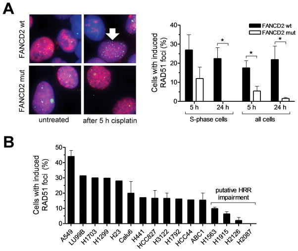
Introduction: Homologous recombination repair (HRR) is a critical pathway for the repair of DNA damage caused by cisplatin or poly-ADP ribose polymerase (PARP) inhibitors. HRR may be impaired by multiple mechanisms in cancer, which complicates assessing the functional HRR status in cells. Here, we monitored the ability of non-small-cell lung cancer (NSCLC) cells to form subnuclear foci of DNA repair proteins as a surrogate of HRR proficiency.

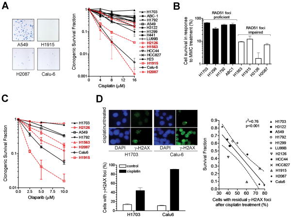
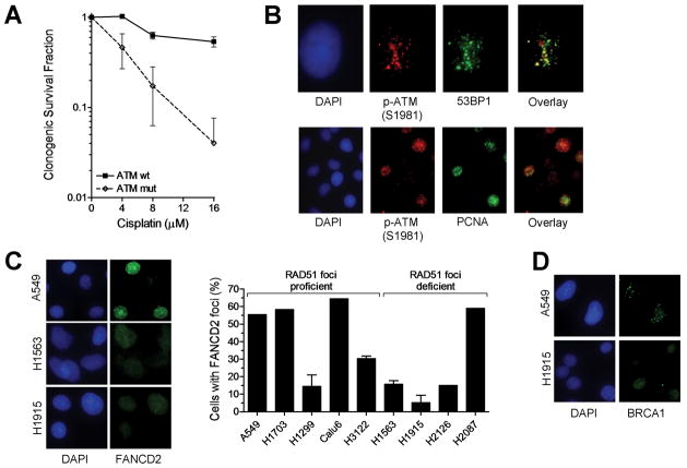
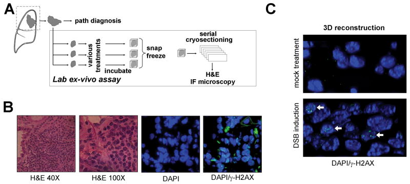
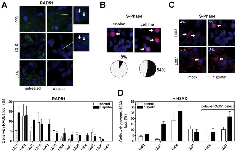
Methods: We assessed clonogenic survival of 16 NSCLC cell lines in response to cisplatin, mitomycin C (MMC), and the PARP inhibitor olaparib. Thirteen tumor explants from patients with NSCLC were subjected to cisplatin ex vivo. Cells were assayed for foci of repair-associated proteins such as BRCA1, FANCD2, RAD51, and γ-H2AX.

Results: Four cell lines (25%) showed an impaired RAD51 foci-forming ability in response to cisplatin. Impaired foci formation correlated with cellular sensitivity to cisplatin, MMC and olaparib. Foci responses complemented or superseded genomic information suggesting alterations in the ATM/ATR and FA/BRCA pathways. Because baseline foci in untreated cells did not predict drug sensitivity, we adapted an ex vivo biomarker assay to monitor damage-induced RAD51 foci in NSCLC explants from patients. Ex vivo cisplatin treatment of explants identified two tumors (15%) exhibiting compromised RAD51 foci induction.

Conclusions: A fraction of NSCLC harbors HRR defects that may sensitize the affected tumors to DNA-damaging agents including PARP inhibitors. We propose that foci-based functional biomarker assays represent a powerful tool for prospective determination of treatment sensitivity, but will require ex vivo techniques for induction of DNA damage to unmask the underlying HRR defect.


Conflict of interest statement

Conflicts of Interest: The authors have no conflicts of interest or relating to the work reported in this manuscript.

Figures

FIGURE 1
FIGURE 1
Monitoring the formation of subnuclear RAD51 foci in response to cisplatin treatment. A, FANCD2-mutant (mut) PD20 fibroblasts with or without exogenous expression of wild-type (wt) FANCD2 were exposed to cisplatin (8 μM) for 1 hour and subjected to immunofluorescence staining for the S-phase marker nuclear PCNA and RAD51 recombinase at 5 and 24 hours (h). Left panel shows representative images of co-staining for PCNA (red) and RAD51 (green), in conjunction with DAPI (blue) counterstaining. Right panel shows fraction of cells with induced RAD51 foci in PCNA-positive S-phase cells or all cells. Bars represent mean with standard error based on 3 independent repeat experiments. Arrow indicates representative nucleus with intact RAD51 foci formation in an S-phase cell. * indicates p<0.05 (t-test, two-tailed). B, Analogous to panel A, 16 NSCLC cell lines were exposed to cisplatin and screened for induced RAD51 foci at 5 h. Bars represent the fraction of cells with at least 15 induced RAD51 foci.
FIGURE 2
FIGURE 2
Sensitivity of NSCLC cell lines to DNA damaging anti-cancer drugs. A, Left panel shows representative examples of colony forming ability after treating A549 cells (200 cells seeded) or cisplatin-sensitive cell lines (2,000-5,000) with 16 μM cisplatin. Right panel, clonogenic survival fraction is plotted against cisplatin concentration. Data points represent mean with standard error based on at least 2 independent repeat experiments. Cell lines with putative RAD51 foci formation defects are highlighted in red. B, Clonogenic survival fraction after treatment with 0.25 mg/ml mitomycin C (MMC) for 1 hour is plotted for a subset of cell lines. C, Clonogenic survival fraction after treatment with 10 μM of olaparib for 72 hours. Cell lines with putative RAD51 foci formation defects are highlighted in red. Bars represent mean with standard error based on at least 2 independent repeat experiments. D, Left panels illustrate the difference in the formation of γ-H2AX foci at 24 hours following treatment of cisplatin-resistant H1703 and cisplatin-sensitive Calu-6 cells with 8 μM cisplatin. Right panel shows the correlation between clonogenic cell survival versus residual γ-H2AX foci present at 24 hours after cisplatin treatment with 8 μM. Dotted line reflects 95% confidence limits of the linear regression line.
FIGURE 3
FIGURE 3
Pathway correlates of cisplatin sensitivity. A, Clonogenic survival of human fibroblasts with wild-type (wt) or mutant (mut) ATM treated with varying doses of cisplatin for 1 hour. Data points represent mean with standard error based on 3 independent repeat experiments. B, Representative images demonstrating co-localization of phospho-ATM foci with 53BP1 or PCNA at 24 hours following cisplatin treatment of A549 cells. C, Analysis of FANCD2 foci formation at 5 hours in a subset of cell lines treated with 8 μM cisplatin. Bars represent the fraction of cells with 20 or more foci per nucleus. D, Representative images illustrating the reduced ability of H1915 cells to form BRCA1 foci in response to cisplatin.
FIGURE 4
FIGURE 4
Ex-vivo foci assay in NSCLC explants. A, Surgical specimens from untreated patients were incubated under standard cell culture conditions and exposed to 8 μM cisplatin, 10 Gy radiation, or mock treatment ex-vivo. Specimens were snap frozen after 5 or 24 hours and subjected to cryosectioning at a later time. B, Serial cryoslides were analyzed by H&E staining to identify viable tumor and immunofluorescence (IF) microscopy to visualize repair protein foci. C, 3-dimensional rendering of tissue nuclei counterstained with γ-H2AX 5 hours after mock treatment or irradiation ex-vivo.
FIGURE 5
FIGURE 5
Detection of cisplatin-induced RAD51 foci in live NSCLC explants. A, Upper panel shows representative images demonstrating subnuclear RAD51 foci following 5 hour incubation after cisplatin. Lower panel displays the fraction of nuclei with RAD51 foci in a panel of 13 tumor explants. Bars represent mean with standard error. B, Illustration of S-phase fraction in representative tumor tissue versus A549 cells using PCNA/DAPI counterstaining (see also Fig. S6C). C, Illustration of PCNA-positive S-phase fraction in RAD51 foci-proficient versus -deficient tumors. D, Quantification of fraction of cells with cisplatin-induced γ-H2AX foci at 5 hours. Bars represent average fraction of nuclei with at least 2 foci +/- standard error based on 8-10 random images and 200-400 nuclei per data point.

References

    1. Shepherd FA, Rosell R. Weighing tumor biology in treatment decisions for patients with non-small cell lung cancer. J Thorac Oncol. 2007;2 (Suppl 2):S68–76. - PubMed
    1. Willers H, Pfäffle HN, Zou L. Targeting Homologous Recombination Repair in Cancer. Academic Press, Elsevier; 2012. pp. 119–160.
    1. Ferrer M, Span SW, Vischioni B, Oudejans JJ, van Diest PJ, de Winter JP, Giaccone G, Kruyt FA. FANCD2 expression in advanced non-small-cell lung cancer and response to platinum-based chemotherapy. Clin Lung Cancer. 2005;6:250–254. - PubMed
    1. Marsit CJ, Liu M, Nelson HH, Posner M, Suzuki M, Kelsey KT. Inactivation of the Fanconi anemia/BRCA pathway in lung and oral cancers: implications for treatment and survival. Oncogene. 2004;23:1000–1004. - PubMed
    1. Rosell R, Skrzypski M, Jassem E, Taron M, Bartolucci R, Sanchez JJ, Mendez P, Chaib I, Perez-Roca L, Szymanowska A, Rzyman W, Puma F, Kobierska-Gulida G, Farabi R, Jassem J. BRCA1: A Novel Prognostic Factor in Resected Non-Small-Cell Lung Cancer. PLoS ONE. 2007;2:e1129. - PMC - PubMed

Publication types

MeSH terms