

Imaging Atherosclerosis by Carotid Intima-media Thickness in vivo: How to, Where and in Whom ?
- PMID: 23399970
- PMCID: PMC3557424
Imaging Atherosclerosis by Carotid Intima-media Thickness in vivo: How to, Where and in Whom ?
Abstract
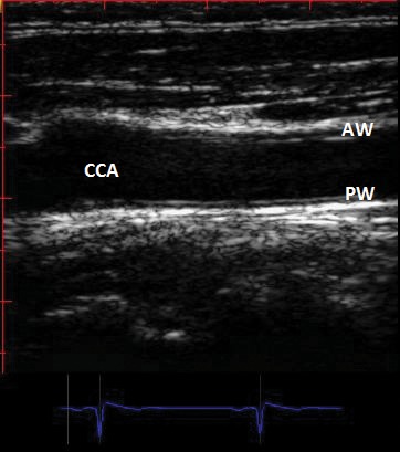
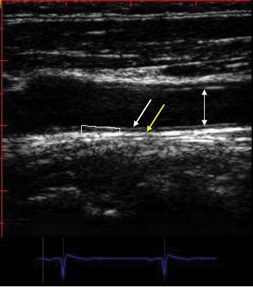
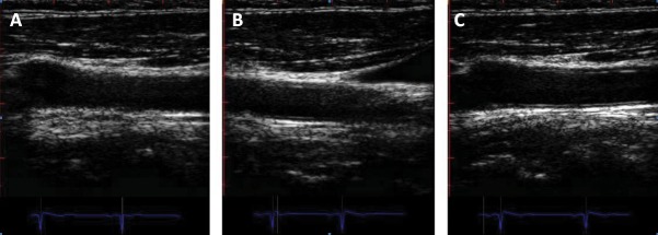
Carotid intima-media thickness (CIMT) can be reliably determined in vivo by carotidian ultrasound and is an accessible and reliable method to assess subclinical atherosclerosis. Available epidemiological data showed that CIMT is significantly correlated with future cardiovascular events. However it has limited value to help risk stratification on top of standard risk-derived functions such as Framingham risk score. It is particularly useful in individuals classified as being at intermediate or high risk by the presence of multiple conventional risk factors.CIMT HAS A CLASS IIA (LOE: B) reccommendation for cardiovascular risk assessment according to the practice guidelines published in 2010, emphasizing the presence of high risk if the common carotid artery intima-media thickness is above the 75(th) percentile. There is no indication to measure IMT in patients with full-blown atherosclerotic carotid disease, although carotidian ultrasound still remains a very useful tool to assess the severity of disease even in these subjects.Progression of CIMT (also associated with increasing age) can be delayed by some drugs (statins, colestipol and niacin) and by risk factors modification. However, there is no consistent data demonstrating a link between progression of CIMT and coronary and cerebral events. Subsequently, studies using CIMT progression as primary outcome to indicate the influence of a certain therapy on cardiovascular risk are inherently misleading as suggested in the recently published ACC/AHA Guidelines.
Keywords: accelerated atherosclerosis; inflammation; metabolic syndrome; rheumatoid arthritis.
Conflict of interest statement
Conflict of interests: We have no conflicts of interest to declare with respect to the authorship of this paper between all and every author and any institution or industry source of funding.
This work is supported by a CNCSIS – UEFISCSU Grant, project number PNII – IDEI, code 257/2008
Figures
References
-
- Ross R. Atherosclerosis: an inflammatory disease. N Engl J Med. 1999;340:115–126. - PubMed
-
- Plutzky J. Inflammatory pathways in atherosclerosis and acute coronary syndromes. Am J Cardiol. 2001;88:10K–15K. - PubMed
-
- Lorenz M. Prediction of clinical cardiovascular events with carotid intima media thickness. A systematic review and meta-analysis. Circulation. 2007;115:459–167. - PubMed
-
- Pignoli P, Tremoli E, Poli A, et al. Intimal plus medial thickness of the arterial wall: a direct measurement with ultrasound imaging. Circulation. 1986;74:1399–1406. - PubMed
-
- O'Leary DH, Bots ML. Imaging of atherosclerosis: carotid intima-media thickness. Eur Heart J. 2010;31:1682–9. - PubMed
LinkOut - more resources
Full Text Sources