. 2014 Oct;18(5):705-12.
doi: 10.1007/s10029-013-1054-2. Epub 2013 Feb 12.

Onlay ventral hernia repairs using porcine non-cross-linked dermal biologic mesh

Affiliations

Onlay ventral hernia repairs using porcine non-cross-linked dermal biologic mesh

E T Alicuben et al. Hernia. 2014 Oct.

Abstract

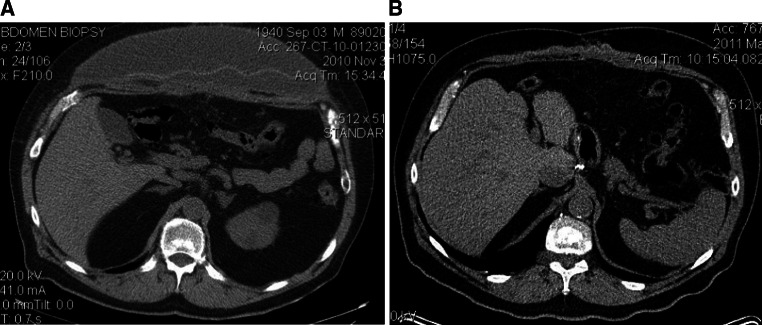
Introduction: Ventral hernias are common and repair with mesh has been shown to reduce recurrence. However, synthetic mesh is associated with a risk of infection. Biologic mesh is an alternative that may be less susceptible to infection. Typically, the sublay position is preferred for mesh placement but this technique takes longer and has not been shown to have a lower recurrence rate than an onlay mesh. The aim of this study was to evaluate the outcome of complex ventral hernia repair using a porcine non-cross-linked biologic mesh onlay.

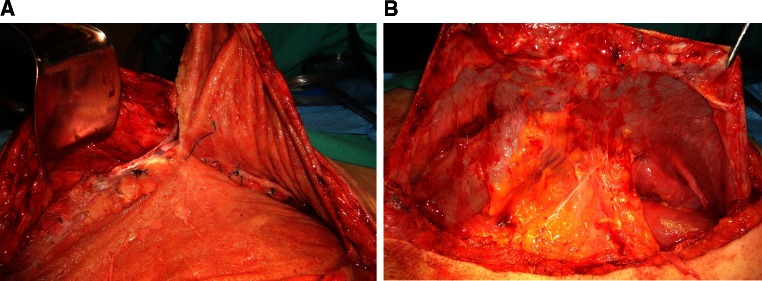
Methods: A retrospective chart review was performed of all patients that had a ventral hernia repair with biologic mesh from January 2009 to March 2012. The operative procedure in all patients was an open repair with primary fascial closure (if possible) with or without external oblique component separation and porcine biologic mesh onlay.

Results: There were 22 patients that had a ventral hernia repair, 19 primary and 3 recurrent. The majority were men, had hernia grade 3 or 4, and developed the hernia after an esophagectomy or gastrectomy for cancer. All but one had primary closure with a porcine biologic mesh onlay. One patient was bridged for loss of domain. A bilateral external oblique component separation was added in 16 patients (73 %). The median hospital stay was 7 days. There were two superficial wound infections, one with exposed mesh, but no patient required mesh removal. A seroma requiring intervention developed in 6 patients (27 %) and resolved with pig-tail drainage. At a median follow-up of 7 months, there has been no hernia recurrence apart from the patient that was bridged.

Conclusions: Porcine non-cross-linked biologic mesh overlay has excellent short-term results in patients at increased risk for mesh infection. No patient required mesh removal, and there have been no recurrent hernias in patients with primary fascial closure. Biologic bridging is not effective for long-term abdominal wall reconstruction.


Figures

Fig. 1
Fig. 1
Completed repair of large ventral hernia. The fascia has been closed in the midline after bilateral external oblique component separations. A 19 × 29 sheet of XenMatrix has been secured in place with #1 proline mattress sutures placed trans-fascially through the mesh circumferentially. Note the trans-fascial sutures are placed lateral to the site of the component separation and the mesh covers this area bilaterally. The midline figure-of-eight proline sutures have also been brought through the mesh to anchor the mesh to the fascia in the midline. The defect was so large that 2 pieces of mesh had to be used and were sewn together. Note the tautness of the mesh. Tension is deliberately placed on the mesh to minimize tension on the midline fascial closure during early healing. The space will be drained with two #10 Jackson-Pratt drains, the deep subcutaneous tissues sewn together and attached to the mesh with 2–0 vicryl, and the skin closed with staples to complete the procedure
Fig. 2
Fig. 2
Slices from an abdominal CT scan showing a large seroma above mesh with portion of mesh in fluid, and b resolution of seroma and intact hernia repair after pig-tail drainage. The mesh is still visible above the fascial closure
Fig. 3
Fig. 3
a Abdominal CT scan and b laparoscopic picture of Spigelian hernia in site where a trans-fascial suture had pulled through the abdominal wall in a patient on large doses of steroids for lung disease that had ventral hernia repair. Note the mesh is still visible on the CT scan after almost a year, and the ventral hernia repair is intact
Fig. 4
Fig. 4
Reoperation 19 months after bridging a patient with loss of domain with XenMatrix. a The mesh has developed into two layers with the subcutaneous tissues incorporated into the superficial part of the mesh. The deeper portion of the mesh is against the viscera. The site of the hernia is just below the retractor where the mesh has separated from the fascia. Note the absence of any form of reconstructed abdominal wall. b The deeper portion of mesh has been separated laterally from the fascia and is being held up by clamps. Neovascularity and adhesions of the omentum and colon to the mesh are visible. The mesh with attached viscera was tucked into the abdomen with no effort to remove it, and since the patient continued to have loss of domain he was bridged with synthetic mesh
Fig. 5
Fig. 5
Re-exploration at 4 months in a patient with cancer. a A full-thickness resection of subcutaneous tissue, XenMatrix mesh, and native abdominal wall (at site of arrow) for histology. Note the incorporation of the subcutaneous tissue (SQ) into the mesh (XM), and the mesh to the abdominal wall (AW). b Histology showing neovascularity going entirely through the mesh from the abdominal wall to the subcutaneous tissue (arrow). c Higher magnification showing infiltration of fibroblasts into the open collagen structure of the mesh (XM)

References

    1. Kingsnorth A, LeBlanc K. Hernias: inguinal and incisional. Lancet. 2003;362:1561–1571. doi: 10.1016/S0140-6736(03)14746-0. - DOI - PubMed
    1. Choi JJ, Palaniappa NC, Dallas KB, et al. Use of mesh during ventral hernia repair in clean-contaminated and contaminated cases: outcomes of 33,832 cases. Ann Surg. 2012;255:176–180. doi: 10.1097/SLA.0b013e31822518e6. - DOI - PubMed
    1. Flum DR, Horvath K, Koepsell T. Have outcomes of incisional hernia repair improved with time? A population-based analysis. Ann Surg. 2003;237:129–135. doi: 10.1097/00000658-200301000-00018. - DOI - PMC - PubMed
    1. Luijendijk RW, Hop WC, van den Tol MP, et al. A comparison of suture repair with mesh repair for incisional hernia. N Engl J Med. 2000;343:392–398. doi: 10.1056/NEJM200008103430603. - DOI - PubMed
    1. Ventral Hernia Working Group. Breuing K, Butler CE, et al. Incisional ventral hernias: review of the literature and recommendations regarding the grading and technique of repair. Surgery. 2010;148:544–558. doi: 10.1016/j.surg.2010.01.008. - DOI - PubMed

Publication types