Periosteal augmentation of allograft bone and its effect on implant fixation - an experimental study on 12 dogs()
- PMID: 23400644
- PMCID: PMC3565231
- DOI: 10.2174/1874325001307010018
Periosteal augmentation of allograft bone and its effect on implant fixation - an experimental study on 12 dogs()
Abstract
Purpose: Periosteum provides essential cellular and biological components necessary for fracture healing and bone repair. We hypothesized that augmenting allograft bone by adding fragmented autologous periosteum would improve fixation of grafted implants.
Methods: In each of twelve dogs, we implanted two unloaded cylindrical (10 mm x 6 mm) titanium implants into the distal femur. The implants were surrounded by a 2.5-mm gap into which morselized allograft bone with or without addition of fragmented autologous periosteum was impacted. After four weeks, the animals were euthanized and the implants were evaluated by histomorphometric analysis and mechanical push-out test.
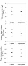
Results: Although less new bone was found on the implant surface and increased volume of fibrous tissue was present in the gap around the implant, no difference was found between treatment groups regarding the mechanical parameters. Increased new bone formation was observed in the immediate vicinity of the periosteum fragments within the bone graft.
Conclusion: The method for periosteal augmentation used in this study did not alter the mechanical fixation although osseointegration was impaired. The observed activity of new bone formation at the boundary of the periosteum fragments may indicate maintained bone stimulating properties of the transplanted cambium layer. Augmenting the bone graft by smaller fragments of periosteum, isolated cambium layer tissue or cultured periosteal cells could be studied in the future.
Keywords: Allograft bone; autologous; implant fixation; morselized; osseointegration; periosteum..
Figures


References
-
- Gie GA, Linder L, Ling RS, et al. Impacted cancellous allografts and cement for revision total hip arthroplasty. J Bone Joint Surg Br . 1993;75(1):14–21. - PubMed
-
- Burchardt H. The biology of bone graft repair. Clin Orthop Relat Res. 1983;(174):28–42. - PubMed
-
- Toms AD, Barker RL, Jones RS, Kuiper JH. Impaction bone-grafting in revision joint replacement surgery. J Bone Joint Surg Am. 2004;86-A(9):2050–60. - PubMed
-
- Van der Donk S, Weernink T, Buma P, et al. Rinsing morselized allografts improves bone and tissue ingrowth. Clin Orthop Relat Res. 2003;(408):302–10. - PubMed
-
- Linder L. Cancellous impaction grafting in the human femur: histological and radiographic observations in 6 autopsy femurs and 8 biopsies. Acta Orthop Scand. 2000;71(6):543–52. - PubMed
Grants and funding
LinkOut - more resources
Full Text Sources
Other Literature Sources
Miscellaneous
