

Efficient Bloch-Siegert B1 (+) mapping using spiral and echo-planar readouts
- PMID: 23401024
- PMCID: PMC3657582
- DOI: 10.1002/mrm.24599
Efficient Bloch-Siegert B1 (+) mapping using spiral and echo-planar readouts
Abstract
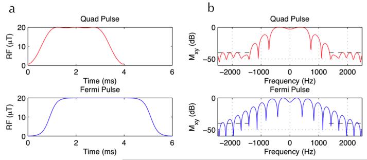
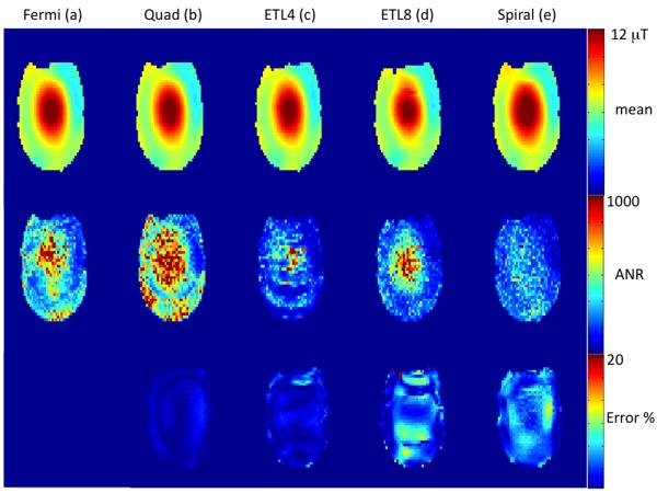
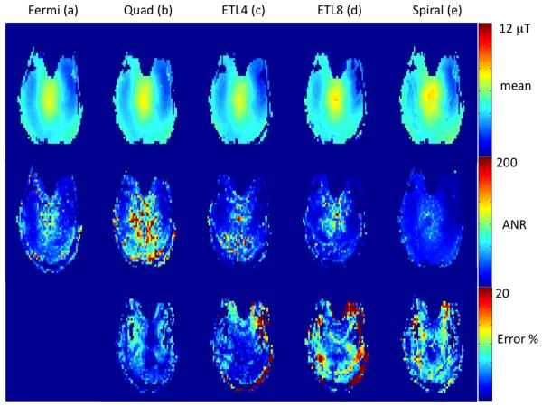
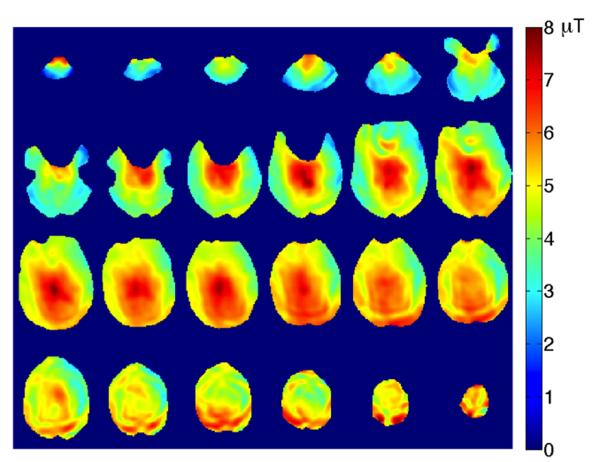
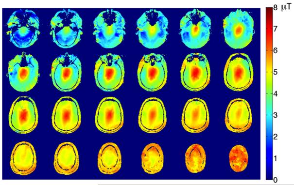
The Bloch-Siegert (B-S) B1 (+) mapping technique is a fast, phase-based method that is highly SAR limited especially at 7T, necessitating the use of long repetition times. Spiral and echo-planar readouts were incorporated in a gradient-echo based B-S sequence to reduce specific absoprtion rate (SAR) and improve its scan efficiency. A novel, numerically optimized 4 ms B-S off-resonant pulse at + 1960 Hz was used to increase sensitivity and further reduce SAR compared with the conventional 6 ms Fermi B-S pulse. Using echo-planar and spiral readouts, scan time reductions of 8-16 were achieved. By reducing the B-S pulse width by a factor of 1.5, SAR was reduced by a factor of 1.5 and overall sensitivity was increased by a factor of 1.33 due to the nearly halved resonance offset of the new B-S pulse. This was validated on phantoms and volunteers at 7 T.
Keywords: B1+ mapping; Bloch-Siegert method; echo-planar readout; spiral readout.
Copyright © 2013 Wiley Periodicals, Inc.
Figures
References
-
- Hornak JP, Szumowski J, Bryant RG. Magnetic field mapping. Magn Reson Med. 1988;6:158–163. - PubMed
-
- Cunningham CH, Pauly JM, Nayak KS. Saturated double-angle method for rapid B1 mapping. Magn Reson Med. 2006;55:1326–1333. - PubMed
-
- Look D, Locker D. Time saving in measurement of NMR and EPR relaxation times. Rev Sci Instrum. 1970;41:250–251.
-
- Wade T, Rutt B. B1 correction using double angle look-locker (DALL). Proceedings of the 16th Annual Meeting of the International Society of Magnetic Resonance in Medicine; Toronto, Canada. 2008. p. p1246.
-
- Yarnykh VL. Actual flip angle imaging in the pulsed steady state: a method for rapid three-dimensional mapping of the transmitted radiofrequency field. Magn Reson Med. 2007;57:192–200. - PubMed
Publication types
MeSH terms
Grants and funding
LinkOut - more resources
Full Text Sources
Other Literature Sources
Miscellaneous
