. 2013 May-Jun;5(3):381-90.
doi: 10.1002/wsbm.1211. Epub 2013 Feb 7.

Mechano-sensing and transduction by endothelial surface glycocalyx: composition, structure, and function

Affiliations

Mechano-sensing and transduction by endothelial surface glycocalyx: composition, structure, and function

Bingmei M Fu et al. Wiley Interdiscip Rev Syst Biol Med. 2013 May-Jun.

Abstract

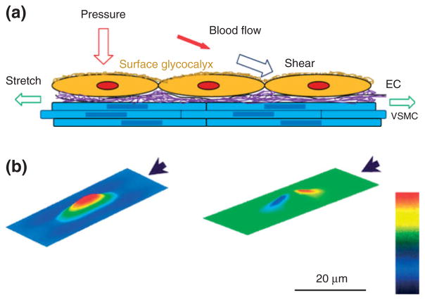
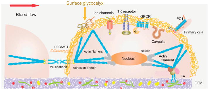
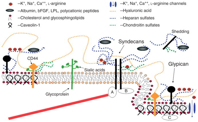
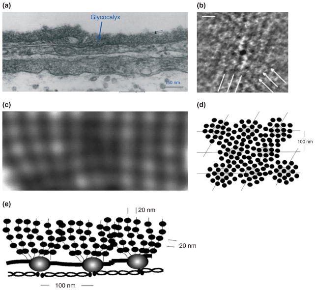
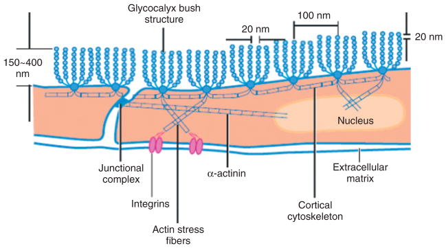
The endothelial cells (ECs) lining every blood vessel wall are constantly exposed to the mechanical forces generated by blood flow. The EC responses to these hemodynamic forces play a critical role in the homeostasis of the circulatory system. To ensure proper EC mechano-sensing and transduction, there are a variety of mechano-sensors and transducers that have been identified on the EC surface, intra- and trans-EC membrane and within the EC cytoskeleton. Among them, the most recent candidate is the endothelial surface glycocalyx (ESG), which is a matrix-like thin layer covering the luminal surface of the EC. It consists of various proteoglycans, glycosaminoglycans, and plasma proteins, and is close to other prominent EC mechano-sensors and transducers. The ESG thickness was found to be in the order of 0.1-1 µm by different visualization techniques and in different types of vessels. Detailed analysis on the electron microscopy (EM) images of the microvascular ESG revealed a quasi-periodic substructure with the ESG fiber diameter of 10-12 and 20 nm spacing between adjacent fibers. Atomic force microscopy and optical tweezers were applied to investigate the mechanical properties of the ESG on the cultured EC monolayers and in solutions. Enzymatic degradation of specific ESG glycosaminoglycan components was used to directly elucidate the role of the ESG in EC mechano-sensing and transduction by measuring the shear-induced productions of nitric oxide and prostacyclin, two characteristic responses of the ECs to the flow. The unique location, composition, and structure of the ESG determine its role in EC mechano-sensing and transduction.


Figures

FIGURE 1
FIGURE 1
(a) Schematic for the forces acting on endothelial cells (ECs) forming the blood vessel wall. Pressure (perpendicular to the ECs) and stretch (in line of the circumferential direction of the vessel wall) due to the blood pressure, shear (tangential to the ECs) due to the blood flow and blood viscosity. Vascular smooth muscle cell (VSMC); purple represents the extra cellular matrix (ECM); at the luminal surface of ECs, there is a thin layer of surface glycocalyx. (b) Predictions from a theoretical model for the distribution of the shear stress (left) and that of the pressure (right) on the surface of the EC. The direction of the flow is represented by the arrow. The bar indicates the magnitude from low (black) to high (red). (Figure 1(b): Reprinted with permission from Ref . Copyright 2004 IOS press)
FIGURE 2
FIGURE 2
Currently identified endothelial mechano-sensors and transducers. At endothelial cell (EC) surface: surface glycocalyx, adherences junction protein VE-cadherin, cell adhesion molecule PECAM-1, ion channels, tyrosine kinase (TK) receptor, G-protein-coupled receptors (GPCR), caveolae, primary cilia, integrins (forming FA, focal adhesion, with ECM protein). In EC cytoskeleton: microtubules and actin filaments. Nesprins connect EC cytoskeleton to nucleus. (Reprinted with permission from Ref . Copright 2011 The Japanese Pharmacological Society)
FIGURE 3
FIGURE 3
A conceptual and simplified view for proteoglycans and glycosaminoglycans (GAGs) of the endothelial surface glycocalyx (ESG). Caveolin-1 associates with regions high in cholesterol and sphingolipids in the EC membrane (darker circles, left), and forms cave-like structures, caveolae (right). Glypicans, along with their HS chains (blue dotted lines) localize in these regions. Transmembrane syndecans are shown to cluster in the outer edge of caveolae. Besides HS, syndecans also contain CS, lower down the core protein (green dotted lines). A glycoprotein with its short oligosaccharide branched chains and their associated SA ‘caps’ are displayed in the middle part of the figure (green). HA is a very long GAG (orange dotted line), which weaves into the glycocalyx and binds with CD44. Transmembrane CD44 can have CS, HS and oligosaccharides attached to it, and localizes in caveolae. Plasma proteins (gray), along with cations and cationic amino acids (red circles) are known to associate with GAGs. (a) The cytoplasmic domains of syndecans can associate with linker molecules which connect them to cytoskeletal elements (red line). (b) Oligomerization of syndecans helps them make direct associations with intracellular signaling effectors. (c) A series of molecules involved in eNOS signaling localize in caveolae. (Reprinted with permission from Ref . Copyright 2006 Blackwell Publishing Ltd)
FIGURE 4
FIGURE 4
EM images and structural model for the endothelial surface glycocalyx (ESG). (a) Electron micrograph of a plastic section of a frog mesenteric capillary. (Reprinted with permission from Ref 47). The thickness of glycocalyx is ~100 nm. (b) Enlarged part of the glycocalyx shown in (a) after processed by autocorrelation functions (ACFs). It shows a quasi-hexagonal arrangement of spacing 80–120 nm. (c) Larger view of glycocalyx showing more detailed structures. (d) En face view (from the luminal side) of a structural model for the glycocalyx corresponding to the EM image shown in (b). (e) Side view of the model for the glycocalyx corresponding to the EM image shown in (c) (Reprinted with permission from Ref . Copyright 2001 Elsevier Science)
FIGURE 5
FIGURE 5
Sketch of a conceptual model for the arrangement of core proteins in the ESG and its anchorage to the underlying actin cortical cytoskeleton. This model is based on the EM observation as shown in Figure 4. (Reprinted with permission from Ref . Copyright 2007 Annual Reviews)

References

    1. Mammoto A, Mammoto T, Ingber DE. Mechanosensitive mechanisms in transcriptional regulation. J Cell Sci. 2012;125(Pt 13):3061–3073. - PMC - PubMed
    1. Davies PF, Dewey CF, Jr, Bussolari SR, Gordon EJ, Gimbrone MA., Jr Influence of hemodynamic forces on vascular endothelial function. In vitro studies of shear stress and pinocytosis in bovine aortic cells. J Clin Invest. 1984;73:1121–1129. - PMC - PubMed
    1. Chien S. Mechanotransduction and endothelial cell homeostasis: the wisdom of the cell. Am J Physiol Heart Circ Physiol. 2007;292:H1209–H224. - PubMed
    1. Ingber DE. Mechanobiology and diseases of mechanotransduction. Ann Med. 2003;35:564–577. - PubMed
    1. Fung YC. Biomechanics: Circulation. New York: Springer; 1997. p. 571.

Publication types

MeSH terms

LinkOut - more resources