. 2013 Mar 11;210(3):563-79.
doi: 10.1084/jem.20120662. Epub 2013 Feb 11.

Deficiency for endoglin in tumor vasculature weakens the endothelial barrier to metastatic dissemination

Affiliations

Deficiency for endoglin in tumor vasculature weakens the endothelial barrier to metastatic dissemination

Charlotte Anderberg et al. J Exp Med. .

Abstract

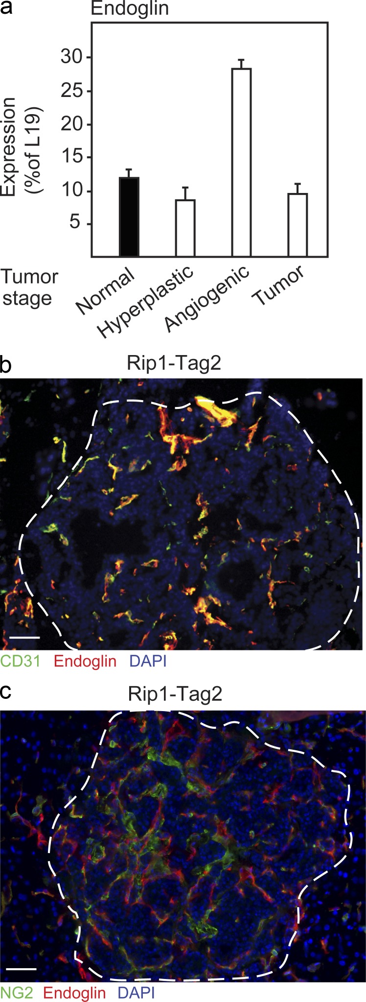
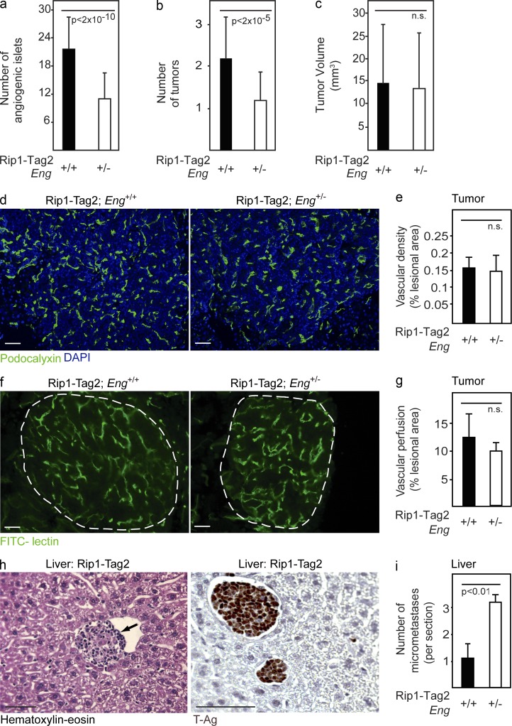
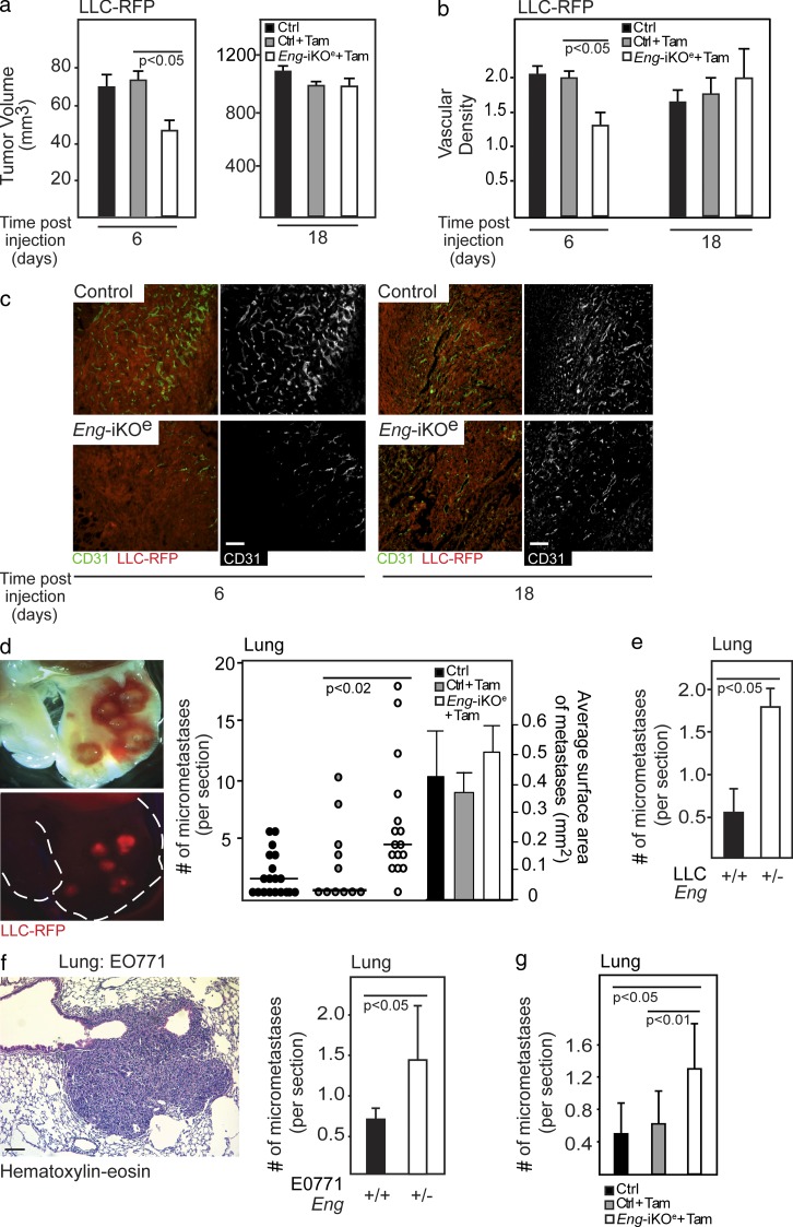
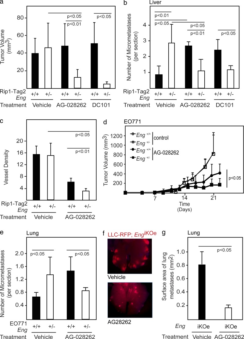
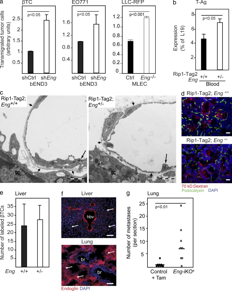
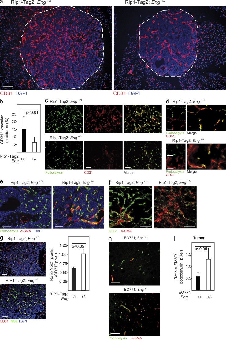
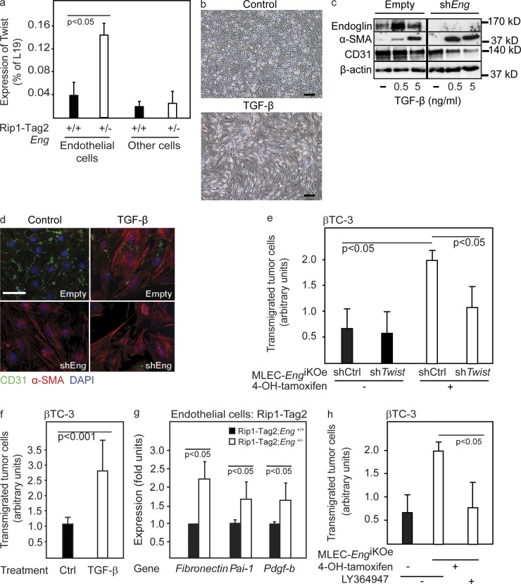
Therapy-induced resistance remains a significant hurdle to achieve long-lasting responses and cures in cancer patients. We investigated the long-term consequences of genetically impaired angiogenesis by engineering multiple tumor models deprived of endoglin, a co-receptor for TGF-β in endothelial cells actively engaged in angiogenesis. Tumors from endoglin-deficient mice adapted to the weakened angiogenic response, and refractoriness to diminished endoglin signaling was accompanied by increased metastatic capability. Mechanistic studies in multiple mouse models of cancer revealed that deficiency for endoglin resulted in a tumor vasculature that displayed hallmarks of endothelial-to-mesenchymal transition, a process of previously unknown significance in cancer biology, but shown by us to be associated with a reduced capacity of the vasculature to avert tumor cell intra- and extravasation. Nevertheless, tumors deprived of endoglin exhibited a delayed onset of resistance to anti-VEGF (vascular endothelial growth factor) agents, illustrating the therapeutic utility of combinatorial targeting of multiple angiogenic pathways for the treatment of cancer.


Figures

Figure 1.
Figure 1.
Endoglin is expressed by endothelial cells during the angiogenic switch of PNET in RIP1-Tag2 mice. (a) Quantitative RT-PCR analysis of expression of endoglin during the different stages of tumorigenic conversion in RIP1-Tag2 mice (a pool of material each derived from >10 mice was used for the analysis). Error bars depict SD. (b) Immunostaining for CD31 and endoglin in PNET lesion from RIP1-Tag2 mice. (c) Immunostaining for the pericyte marker NG2 and endoglin in PNET lesion from RIP1-Tag2 mice. (b and c) Cell nuclei are shown with DAPI. The dashed line marks the tumor-exocrine pancreas boundary. Bars, 50 µm.
Figure 2.
Figure 2.
Increased metastatic seeding of tumors in endoglin-deficient mice. (a–c) Quantification of number of angiogenic islets (a), number of tumors (b), and total tumor burden (c) in 12-wk-old RIP1-Tag2;Eng+/+ (n = 22) and RIP1-Tag2;Eng+/− mice (n = 55). (d and e) Immunostaining (d) and quantification (e) of total positively stained area for the vascular marker podocalyxin in PNETs from RIP1-Tag2;Eng+/+ and RIP1-Tag2;Eng+/− mice. Analysis was performed using at least five mice per group. (f and g) Perfused vessels in PNETs from RIP1-Tag2;Eng+/+ and RIP1-Tag2;Eng+/− mice visualized (f) and quantified (g) by fluorescein-labeled tomato lectin. The dashed line marks the tumor-exocrine pancreas boundary. Analysis was performed using three mice per group. (d and f) Cell nuclei are shown with DAPI. (h) Representative picture of hematoxylin-eosin staining (left) and immunostaining for SV40 T-antigen (T-Ag; right) of a micrometastatic liver lesion. The arrow inside the sinusoidal vessel marks metastatic lesion. (i) Quantification of micrometastatic foci in the liver of RIP1-Tag2;Eng+/+ and RIP1-Tag2;Eng+/− mice at 12 wk of age. Error bars depict SD; n.s., not significantly different. Bars, 50 µm.
Figure 3.
Figure 3.
Increased metastatic seeding in endoglin-deficient mice is an endothelial cell–autonomous event. (a) In vivo tumor volume of subcutaneously injected LLC-RFP cells at 6 and 18 d in control (n = 7 and n = 19, respectively), tamoxifen-treated control (n = 7 and n = 11, respectively), and EngiKOe (n = 9 and n = 15, respectively) mice. (b and c) Quantification (b) and immunostaining (c) of vessel density of LLC-RFP tumors using CD31 during initial phases of tumor development and at the endpoint of the experiment in control (n = 7 and n = 19, respectively), tamoxifen-treated controls (n = 7 and n = 11, respectively), and EngiKOe (n = 9 and n = 15, respectively) mice. (d) Representative picture and quantification of lung metastases using fluorescent imaging of RFP-tagged LLC cells (LLC-RFP) in control (n = 19), tamoxifen-treated control (n = 11) or EngiKOe (n = 15) mice with established subcutaneous tumors. A subset of mice (n = 8, 6, and 6, respectively) was also analyzed for the mean size of metastatic lesions. The horizontal lines represent median. Statistical analysis was performed using the Mann-Whitney U test. (e) Quantification of lung metastases of LLC-RFP tumors established subcutaneously in Eng+/+ (n = 11) or Eng+/− (n = 9) mice. (f) Representative picture of hematoxylin-eosin staining of a micrometastatic lung lesion from EO771 tumors. Quantification of micrometastatic foci in the lung of EO771-bearing Eng+/+ (n = 5) and Eng+/− (n = 5) mice. (g) Quantification of lung metastases from EO771 tumors established subcutaneously in control (n = 8), tamoxifen-treated control (Tam; n = 8), or EngiKOe (n = 8) mice. Error bars depict SD. Bars, 50 µm.
Figure 4.
Figure 4.
Endoglin deficiency maintains tumors in a sensitive state to VEGF inhibition. (a) Total tumor burden of RIP1-Tag2;Eng+/+ and RIP1-Tag2;Eng+/− mice at 13 wk of age after 3 wk of treatment with vehicle (n = 6 and n = 5, respectively), the small molecule VEGFR inhibitor AG-028262 (n = 9 and n = 5, respectively), or the VEGFR2-neutralizing antibody DC101 (n = 3 in each group). The data shown are representative of two independent experiments. (b) Quantification of micrometastatic PNET foci in the liver of RIP1-Tag2;Eng+/+ and RIP1-Tag2;Eng+/− mice at 13 wk of age after 3 wk of treatment with vehicle (n = 4 in each group), AG-028262 (n = 4 in each group), or DC101 (n = 3 in each group). (c) Quantification of total positively stained area for the vascular marker podocalyxin in PNET from RIP1-Tag2;Eng+/+ and RIP1-Tag2;Eng+/− mice after 3 wk treatment with vehicle or AG-028262. (d and e) In vivo growth rate (d) and quantification of micrometastatic lung foci (e) of orthotopically implanted EO771 mammary tumors in Eng+/+ and Eng+/− mice during treatment with control or AG-028262 (n = 5–11). Data from control mice are reproduced from Fig. 3 f to allow easy comparison. (f and g) Representative image (f) and quantification (g) of metastatic foci of LLC-RFP tumors in the lungs of EngiKOe mice treated with vehicle or AG-028262. Error bars depict SD.
Figure 5.
Figure 5.
The increased metastatic seeding in endoglin-deficient mice is a result of disruption of the endothelial cell barrier. (a) Transmigration of βTC-3 cells derived from a PNET in a RIP1-Tag2 mouse, EO771 cells, or LLC-RFP cells through a monolayer of bEnd3.1 endothelial cells expressing either shRNA-endoglin or control vector or through a monolayer of primary mouse lung endothelial cells isolated from EngiKOe mice carrying the Immortomouse transgene (MLEC). The mean of two to three independent experiments is shown. (b) Quantitative RT-PCR detection of SV40 T-Ag transcripts in whole blood from RIP1-Tag2;Eng+/+ (n = 10) and RIP1-Tag2;Eng+/− (n = 5) mice. (c) Electron micrographic views of transversely cut tumor microvessels in RIP1-Tag2;Eng+/+ and RIP1-Tag2;Eng+/− mice. Interendothelial junctions are indicated from the luminal aspect (long arrows). Continuous periendothelial basal laminae (short arrows) invest cytoplasmic strands of pericytes (white asterisks). tc, tumor cell. (d) Detection of extravasated fluorescently labeled 70 kD dextran and immunostaining for podocalyxin of PNETs from RIP1-Tag2;Eng+/+ mice and RIP1-Tag2;Eng+/− excised 4 min after injection of dextran. (e) Quantification of extravasated βTC-3 cell labeled with a fluorescent tracer (green) in the liver parenchyma from Eng+/+ (n = 4) and Eng+/− (n = 3) mice 24 h after tail vein injection. (f) Immunohistochemical detection of endoglin in liver sinusoidal endothelial cells and lung capillaries. Cell nuclei are shown with DAPI. Arrows point out sinusoidal and lung capillaries. hbv, hepatic blood vessel; br, bronchiolus. (g) Quantification of LLC-RFP metastatic foci in the lung 15 d after tail vein injection in WT tamoxifen-treated (n = 9) and EngiKOe (n = 8). Horizontal lines represent median. Statistical analysis was performed using the Mann-Whitney U test. Error bars depict SD. Bars: (c) 1 µm; (d and f) 50 µm.
Figure 6.
Figure 6.
Reduced endoglin expression gives rise to hallmarks of EndMT in tumors in vivo. (a) Immunostaining for CD31 of PNETs from 12-wk-old RIP1-Tag2;Eng+/+ and RIP1-Tag2;Eng+/− mice. The dashed line marks the tumor-exocrine pancreas boundary. (b) Quantification of vessels with high CD31 expression in PNET lesions from RIP1-Tag2;Eng+/+ (n = 3) and RIP1-Tag2;Eng+/− (n = 4) mice. (c and d) Immunostaining for podocalyxin and CD31 of PNET lesions from 12-wk-old RIP1-Tag2;Eng+/+ and RIP1-Tag2;Eng+/− mice. (e) Immunostaining for podocalyxin and α-SMA of PNET lesions from RIP1-Tag2;Eng+/+ and RIP1-Tag2;Eng+/− mice. (f) Representative confocal micrograph of immunostaining for CD31 and α-SMA of PNET lesion from 12-wk-old RIP1-Tag2;Eng+/+ and RIP1-Tag2;Eng+/− mice. (g) Immunostaining and quantification of NG2 and CD31 of sections of PNET from 12-wk-old RIP1-Tag2 mice. (a, e, and g) Cell nuclei are shown with DAPI. (h) Immunostaining for podocalyxin and α-SMA of EO771 tumors grown in Eng+/+ or Eng+/− mice.(i) Quantification of α-SMA+ vessels in EO771 tumors from Eng+/+ (n = 4) and Eng+/− (n = 4) mice. Error bars depict SD. Bars: (a) Bars, 100 µm; (c–h) 50 µm.
Figure 7.
Figure 7.
Reduced expression of endoglin in endothelial cells facilitates EndMT and tumor cell transmigration. (a) Quantitative RT-PCR for Twist in isolated endothelial cells or nonendothelial cells (other cells) from PNETs of 12-wk-old RIP1-Tag2;Eng+/+ and RIP1-Tag2;Eng+/− mice. A representative experiment using a pool of material derived from more than six mice per group is shown. (b) Micrograph of bEnd3.1 endothelial cells stimulated with TGF-β for 48 h. (c) Western blot of endoglin, α-SMA, and CD31 in bEnd3.1 cells expressing either shRNA-endoglin or empty vector stimulated with TGF-β. β-Actin served as a loading control. The dividing line marks duplicate lanes that were removed. (d) Immunostaining for CD31 and α-SMA of bEnd3.1/shRNA-endoglin or control cells with and without TGF-β stimulation. (e) Transmigration of βTC-3 cells through a monolayer of MLEC either induced or noninduced to knock out the gene for endoglin by treatment with 4-OH-tamoxifen and infected with a lentiviral control construct (shCtrl) or shTwist construct. The mean of two independent experiments is shown. (f) Transmigration of βTC-3 cells through a monolayer of MS1 pancreatic islet endothelial cells stimulated with TGF-β. The mean of three independent experiments is shown. (g) Quantitative RT-PCR for the ALK5 target genes fibronectin (Fn), plasminogen activator inhibitor-1 (PAI-1), and platelet-derived growth factor B (PDGF-B). A pool of endothelial cell–specific genes (VE-cadherin, CD31, and VEGFR-2) was used as a reference. The mean of two independent experiments is shown (a pool of material derived from more than six mice per group was used for the analysis). (h) Transmigration of β-TC3 cells through a monolayer of MLEC either induced or noninduced to knock out the gene for endoglin by treatment with 4-OH-tamoxifen and in the presence of 1 µM of the ALK5 inhibitor LY364947. The mean of two independent experiments is shown. Error bars depict SD. Bars: (b) 50 µm; (d) 20 µm.

References

    1. Arthur H.M., Ure J., Smith A.J., Renforth G., Wilson D.I., Torsney E., Charlton R., Parums D.V., Jowett T., Marchuk D.A., et al. 2000. Endoglin, an ancillary TGFbeta receptor, is required for extraembryonic angiogenesis and plays a key role in heart development. Dev. Biol. 217:42–53 10.1006/dbio.1999.9534 - DOI - PubMed
    1. Bergers G., Hanahan D. 2008. Modes of resistance to anti-angiogenic therapy. Nat. Rev. Cancer. 8:592–603 10.1038/nrc2442 - DOI - PMC - PubMed
    1. Bernabeu C., Lopez-Novoa J.M., Quintanilla M. 2009. The emerging role of TGF-beta superfamily coreceptors in cancer. Biochim. Biophys. Acta. 1792:954–973 10.1016/j.bbadis.2009.07.003 - DOI - PubMed
    1. Blanco F.J., Santibanez J.F., Guerrero-Esteo M., Langa C., Vary C.P., Bernabeu C. 2005. Interaction and functional interplay between endoglin and ALK-1, two components of the endothelial transforming growth factor-beta receptor complex. J. Cell. Physiol. 204:574–584 10.1002/jcp.20311 - DOI - PubMed
    1. Bourdeau A., Dumont D.J., Letarte M. 1999. A murine model of hereditary hemorrhagic telangiectasia. J. Clin. Invest. 104:1343–1351 10.1172/JCI8088 - DOI - PMC - PubMed

Publication types

MeSH terms

Substances