Review
. 2013 May;34(17):1270-8.
doi: 10.1093/eurheartj/eht023. Epub 2013 Feb 10.

The eye and the heart

Affiliations
Review

The eye and the heart

Josef Flammer et al. Eur Heart J. 2013 May.

Abstract

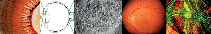
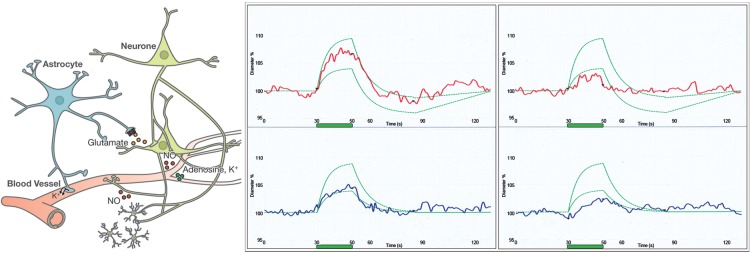
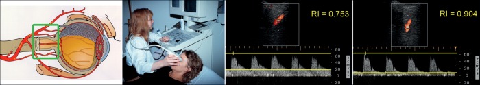
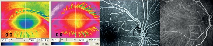
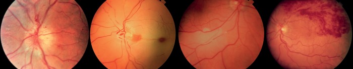
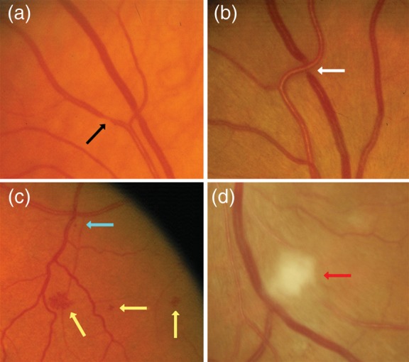
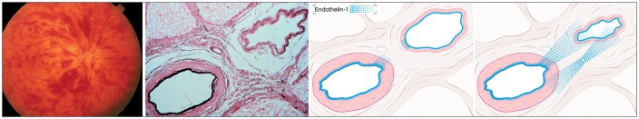
The vasculature of the eye and the heart share several common characteristics. The easily accessible vessels of the eye are therefore-to some extent-a window to the heart. There is interplay between cardiovascular functions and risk factors and the occurrence and progression of many eye diseases. In particular, arteriovenous nipping, narrowing of retinal arteries, and the dilatation of retinal veins are important signs of increased cardiovascular risk. The pressure in the dilated veins is often markedly increased due to a dysregulation of venous outflow from the eye. Besides such morphological criteria, functional alterations might be even more relevant and may play an important role in future diagnostics. Via neurovascular coupling, flickering light dilates capillaries and small arterioles, thus inducing endothelium-dependent, flow-mediated dilation of larger retinal vessels. Risk factors for arteriosclerosis, such as dyslipidaemia, diabetes, or systemic hypertension, are also risk factors for eye diseases such as retinal arterial or retinal vein occlusions, cataracts, age-related macular degeneration, and increases in intraocular pressure (IOP). Functional alterations of blood flow are particularly relevant to the eye. The primary vascular dysregulation syndrome (PVD), which often includes systemic hypotension, is associated with disturbed autoregulation of ocular blood flow (OBF). Fluctuation of IOP on a high level or blood pressure on a low level leads to instable OBF and oxygen supply and therefore to oxidative stress, which is particularly involved in the pathogenesis of glaucomatous neuropathy. Vascular dysregulation also leads to a barrier dysfunction and thereby to small retinal haemorrhages.

Keywords: Cardiovascular risk; Endothelial function; Glaucoma; Retinal vein occlusion; Retinal venous pressure; Retinal vessels; Systemic hypertension; Systemic hypotension; Vascular dysregulation.


Figures

Figure 1
Figure 1
The ciliary body is highly perfused and produces the aqueous humour (left: photo taken from the back of the eye). The optic nerve head has a very dense network of long capillaries (middle). The retinal circulation is similar to brain circulation but without autonomic innervation. In contrast, the vasculature of the choroid is densely innervated (right).
Figure 2
Figure 2
The size of the retinal vessels is influenced by neural and glial cells (neurovascular coupling), shown in a simplified view on the left. Flickering light (green bar) leads to vasodilation of arteries (red) and veins (blue) in healthy subjects (middle) and to a lesser extent in subjects with vascular dysregulation (right). The green curves indicate the normal range. (Modified after Flammer J, Mozaffarieh M, Bebie H. Basic Sciences in Ophthalmology–Physics and Chemistry. Springer Publications, in print, with permission.)
Figure 3
Figure 3
The vessels behind the eye (ophthalmic artery, central retinal artery, and the ciliary arteries) can be visualized and its flow quantified by colour Doppler imaging. Shown is the outcome from the ophthalmic artery of a healthy subject with normal resistivity (middle) and of a glaucoma patient with high resistivity (right). (Modified after Flammer J, Mozaffarieh M, Bebie H. Basic Sciences in Ophthalmology–Physics and Chemistry. Springer Publications, in print, with permission.)
Figure 4
Figure 4
The bulk flow can be quantified with the help of thermography. Left: A relatively cool eye of a subject with vascular dysregulation in relation to a normal control (middle left). The retinal circulation is visualized with fluorescence angiography (middle right) and choroid circulation with the indocyanine green angiography (right).
Figure 5
Figure 5
Classical ocular blood flow dysfunctions: (i) Anterior ischaemic neuropathy. (ii) Central retinal arterial occlusion. (iii) Embolus in a retinal artery. (iv) Retinal branch vein occlusion.
Figure 6
Figure 6
Examples of retinal vascular signs in patients with cardiovascular diseases. Black arrow: focal arteriolar narrowing. White arrow: arterio-venous nicking. Yellow arrow: haemorrhage. Blue arrow: micro-aneurysm. Red arrow: cotton wool spot. (From Liew and Wang, reused with permission from the author and the publisher.)
Figure 7
Figure 7
Left: Under hypoxic condition hypoxia-inducible factor-1 alpha (HIF-1α) is increased and enhances expression of genes such as endothelin-1 or vascular endothelial growth factor. (From Flammer J, Mozaffarieh M, Bebie H. Basic Sciences in Ophthalmology–Physics and Chemistry. Springer Publications, in print, with permission.) This leads to weakening of the BRB (an example is the macular oedema, second from left) or to neovascularization (an example is wet age-related macular degeneration, second from right) Right: Antibody or antibody fragment injection into the eye binds VEGF, thereby restoring wet age-related macular degeneration in a dry age-related macular degeneration.
Figure 8
Figure 8
In the optic nerve head (ONH) (second from left), the blood–brain barrier is partly abrogated by the proximity to the fenestrated vessels of the choroid (left). Unstable oxygen supply in glaucoma patients increases superoxide anion (O2) in the mitochondria of the axons. If neighbouring astrocytes are activated, nitric oxide (NO) diffuses into the axons resulting in the damaging peroxynitrite (ONOO) (second from right). Indeed, visual field progression in glaucoma patients (right) increases not only with increasing intraocular pressure (green) but also with decreasing ocular blood flow (red). (From Flammer and Mozaffarieh, with permission.)
Figure 9
Figure 9
Pathogenesis of optic disc splinter haemorrhages: Under normal conditions, the vessels in and around the optic nerve head are watertight. If the barrier is opened at the level of the endothelial cells, small molecules such as water as well as fluorescein can leak out. If, at the same time, the basal membrane in the same area is also weakened, erythrocytes can also escape. (Modified after Grieshaber and Flammer, with permission.)
Figure 10
Figure 10
Pathogenesis of retinal vein occlusion: At the lamina cribrosa, the central artery and central vein are topographically very close and share a common adventitia (middle). This enables a molecular cross talk between the two vessels (right). Endothelin-1 (blue), for example, can diffuse from the ailing artery as well as from the adjacent hypoxic tissue to the very sensitive vein, leading to venous constriction. [Modified after Fraenkl SA, Mozaffarieh M, Flammer J. (2010), Figures 1a, 2, 4). With kind permission from Springer Science + Business Media B.V.]

References

    1. Mozaffarieh M, Flammer J. Ocular Blood Flow and Glaucomatous Optic Neuropathy. 1st ed. Berlin/Heidelberg: Springer; 2009.
    1. Flammer J, Mozaffarieh M. Autoregulation, a balancing act between supply and demand. Can J Ophthalmol. 2008;43:317–321. - PubMed
    1. Kur J, Newman EA, Chan-Ling T. Cellular and physiological mechanisms underlying blood flow regulation in the retina and choroid in health and disease. Prog Retin Eye Res. 2012;31:377–406. - PMC - PubMed
    1. Kotliar KE, Mucke B, Vilser W, Schilling R, Lanzl IM. Effect of aging on retinal artery blood column diameter measured along the vessel axis. Invest Ophthalmol Vis Sci. 2008;49:2094–2102. - PubMed
    1. Flammer AJ, Anderson T, Celermajer DS, Creager MA, Deanfield J, Ganz P, Hamburg NM, Luscher TF, Shechter M, Taddei S, Vita JA, Lerman A. The assessment of endothelial function: from research into clinical practice. Circulation. 2012;126:753–767. - PMC - PubMed