Severity of cardiomyopathy associated with adenine nucleotide translocator-1 deficiency correlates with mtDNA haplogroup
- PMID: 23401503
- PMCID: PMC3587196
- DOI: 10.1073/pnas.1300690110
Severity of cardiomyopathy associated with adenine nucleotide translocator-1 deficiency correlates with mtDNA haplogroup
Abstract
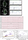
Mutations of both nuclear and mitochondrial DNA (mtDNA)-encoded mitochondrial proteins can cause cardiomyopathy associated with mitochondrial dysfunction. Hence, the cardiac phenotype of nuclear DNA mitochondrial mutations might be modulated by mtDNA variation. We studied a 13-generation Mennonite pedigree with autosomal recessive myopathy and cardiomyopathy due to an SLC25A4 frameshift null mutation (c.523delC, p.Q175RfsX38), which codes for the heart-muscle isoform of the adenine nucleotide translocator-1. Ten homozygous null (adenine nucleotide translocator-1(-/-)) patients monitored over a median of 6 years had a phenotype of progressive myocardial thickening, hyperalaninemia, lactic acidosis, exercise intolerance, and persistent adrenergic activation. Electrocardiography and echocardiography with velocity vector imaging revealed abnormal contractile mechanics, myocardial repolarization abnormalities, and impaired left ventricular relaxation. End-stage heart disease was characterized by massive, symmetric, concentric cardiac hypertrophy; widespread cardiomyocyte degeneration; overabundant and structurally abnormal mitochondria; extensive subendocardial interstitial fibrosis; and marked hypertrophy of arteriolar smooth muscle. Substantial variability in the progression and severity of heart disease segregated with maternal lineage, and sequencing of mtDNA from five maternal lineages revealed two major European haplogroups, U and H. Patients with the haplogroup U mtDNAs had more rapid and severe cardiomyopathy than those with haplogroup H.
Conflict of interest statement
The authors declare no conflict of interest.
Figures


Similar articles
-
Complete loss-of-function of the heart/muscle-specific adenine nucleotide translocator is associated with mitochondrial myopathy and cardiomyopathy.Hum Mol Genet. 2005 Oct 15;14(20):3079-88. doi: 10.1093/hmg/ddi341. Epub 2005 Sep 9. Hum Mol Genet. 2005. PMID: 16155110
-
Mitochondrial DNA Variation Dictates Expressivity and Progression of Nuclear DNA Mutations Causing Cardiomyopathy.Cell Metab. 2019 Jan 8;29(1):78-90.e5. doi: 10.1016/j.cmet.2018.08.002. Epub 2018 Aug 30. Cell Metab. 2019. PMID: 30174309 Free PMC article.
-
Mitochondrial cardiomyopathies: how to identify candidate pathogenic mutations by mitochondrial DNA sequencing, MITOMASTER and phylogeny.Eur J Hum Genet. 2011 Feb;19(2):200-7. doi: 10.1038/ejhg.2010.169. Epub 2010 Oct 27. Eur J Hum Genet. 2011. PMID: 20978534 Free PMC article.
-
Two new cases of mitochondrial myopathy with exercise intolerance, hyperlactatemia and cardiomyopathy, caused by recessive SLC25A4 mutations.Mitochondrion. 2018 Mar;39:26-29. doi: 10.1016/j.mito.2017.08.009. Epub 2017 Aug 18. Mitochondrion. 2018. PMID: 28823815 Review.
-
The adenine nucleotide translocase type 1 (ANT1): a new factor in mitochondrial disease.IUBMB Life. 2005 Sep;57(9):607-14. doi: 10.1080/15216540500217735. IUBMB Life. 2005. PMID: 16203679 Review.
Cited by
-
NME6 is a phosphotransfer-inactive, monomeric NME/NDPK family member and functions in complexes at the interface of mitochondrial inner membrane and matrix.Cell Biosci. 2021 Nov 17;11(1):195. doi: 10.1186/s13578-021-00707-0. Cell Biosci. 2021. PMID: 34789336 Free PMC article.
-
Mitochondrial DNA genetics and the heteroplasmy conundrum in evolution and disease.Cold Spring Harb Perspect Biol. 2013 Nov 1;5(11):a021220. doi: 10.1101/cshperspect.a021220. Cold Spring Harb Perspect Biol. 2013. PMID: 24186072 Free PMC article. Review.
-
Inefficient Batteries in Heart Failure: Metabolic Bottlenecks Disrupting the Mitochondrial Ecosystem.JACC Basic Transl Sci. 2022 Jul 20;7(11):1161-1179. doi: 10.1016/j.jacbts.2022.03.017. eCollection 2022 Nov. JACC Basic Transl Sci. 2022. PMID: 36687274 Free PMC article. Review.
-
The genetic landscape of cardiomyopathy and its role in heart failure.Cell Metab. 2015 Feb 3;21(2):174-182. doi: 10.1016/j.cmet.2015.01.013. Cell Metab. 2015. PMID: 25651172 Free PMC article. Review.
-
Risks inherent to mitochondrial replacement.EMBO Rep. 2015 May;16(5):541-4. doi: 10.15252/embr.201439110. Epub 2015 Mar 25. EMBO Rep. 2015. PMID: 25807984 Free PMC article. No abstract available.
References
-
- Nishizawa M, et al. A mitochondrial encephalomyopathy with cardiomyopathy. A case revealing a defect of complex I in the respiratory chain. J Neurol Sci. 1987;78(2):189–201. - PubMed
-
- Wallace DC, Lott MT, Procaccio V. 2013. Emery and Rimoin’s Principles and Practice of Medical Genetics, Mitochondrial Medicine: The Mitochondrial Biology and Genetics of Metabolic and Degenerative Diseases, Cancer, and Aging, Chapter 13, eds Rimoin DL, Pyeritz RE, Korf BR (Churchill Livingstone Elsevier, Philadelphia), 6th Ed, Vol 1.
-
- Palmieri L, et al. Complete loss-of-function of the heart/muscle-specific adenine nucleotide translocator is associated with mitochondrial myopathy and cardiomyopathy. Hum Mol Genet. 2005;14(20):3079–3088. - PubMed
Publication types
MeSH terms
Substances
Grants and funding
LinkOut - more resources
Full Text Sources
Other Literature Sources
Medical
Molecular Biology Databases
Research Materials

