. 2013 Feb 26;110(9):3513-8.
doi: 10.1073/pnas.1214099110. Epub 2013 Feb 11.

Alternative erythropoietin-mediated signaling prevents secondary microvascular thrombosis and inflammation within cutaneous burns

Affiliations

Alternative erythropoietin-mediated signaling prevents secondary microvascular thrombosis and inflammation within cutaneous burns

Stefan Bohr et al. Proc Natl Acad Sci U S A. .

Abstract

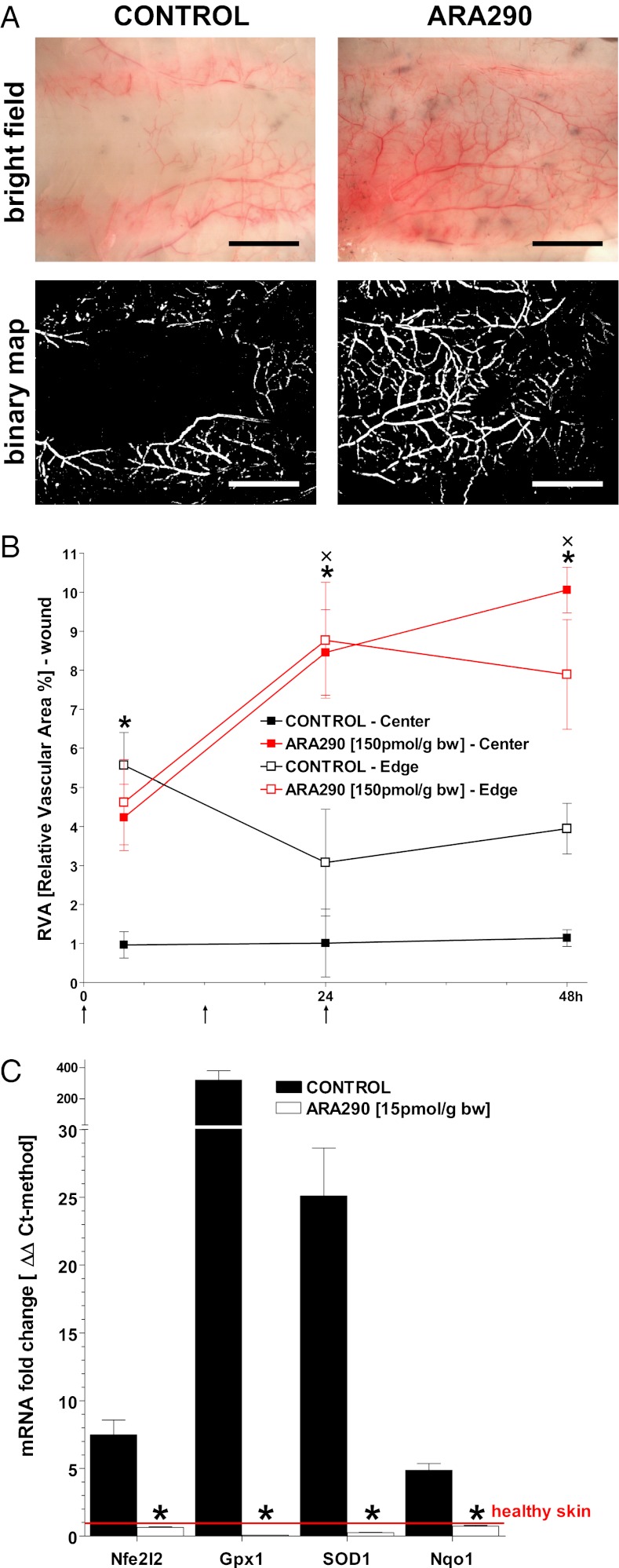
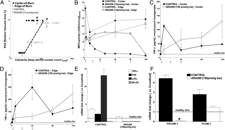
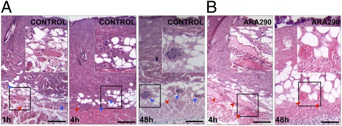
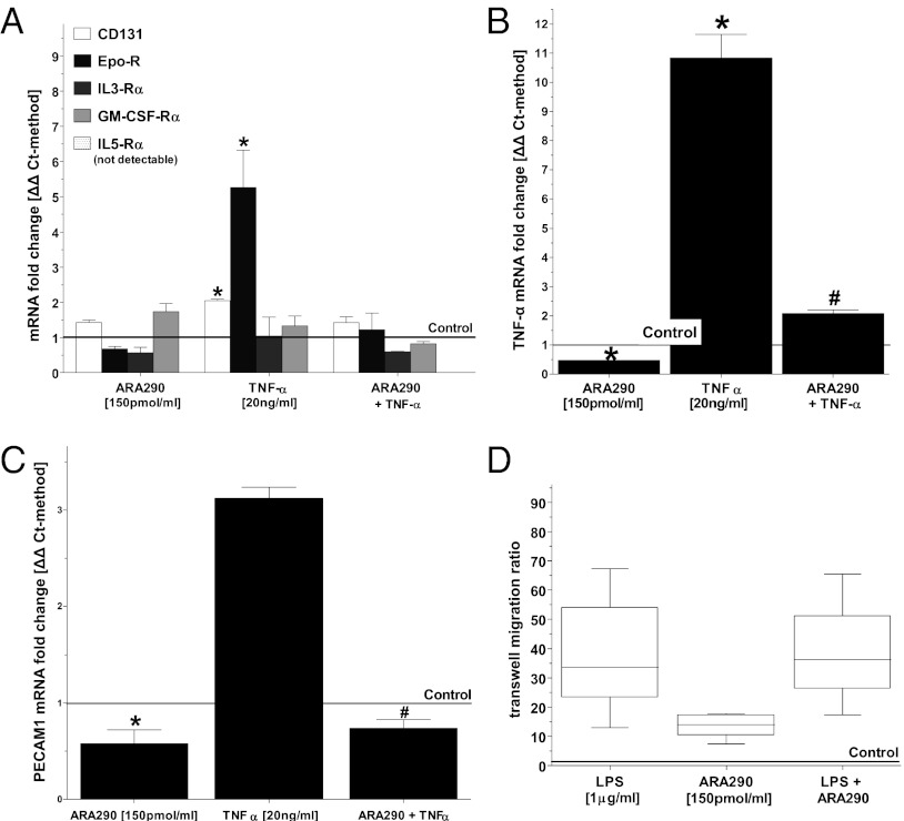
Alternate erythropoietin (EPO)-mediated signaling via the heteromeric receptor composed of the EPO receptor and the β-common receptor (CD131) exerts the tissue-protective actions of EPO in various types of injuries. Herein we investigated the effects of the EPO derivative helix beta surface peptide (synonym: ARA290), which specifically triggers alternate EPO-mediated signaling, but does not bind the erythropoietic EPO receptor homodimer, on the progression of secondary tissue damage following cutaneous burns. For this purpose, a deep partial thickness cutaneous burn injury was applied on the back of mice, followed by systemic administration of vehicle or ARA290 at 1, 12, and 24 h postburn. With vehicle-only treatment, wounds exhibited secondary microvascular thrombosis within 24 h postburn, and subsequent necrosis of the surrounding tissue, thus converting to a full-thickness injury within 48 h. On the other hand, when ARA290 was systemically administered, patency of the microvasculature was maintained. Furthermore, ARA290 mitigated the innate inflammatory response, most notably tumor necrosis factor-alpha-mediated signaling. These findings correlated with long-term recovery of initially injured yet viable tissue components. In conclusion, ARA290 may be a promising therapeutic approach to prevent the conversion of partial- to full-thickness burn injuries. In a clinical setting, the decrease in burn depth and area would likely reduce the necessity for extensive surgical debridement as well as secondary wound closure by means of skin grafting. This use of ARA290 is consistent with its tissue-protective properties previously reported in other models of injury, such as myocardial infarction and hemorrhagic shock.


Conflict of interest statement

Conflict of interest statement: M.B. and A.C. are officers of Araim Pharmaceuticals and currently hold stock in the company.

Figures

Fig. 1.
Fig. 1.
ARA290 treatment salvages microvascular perfusion throughout wound beds. (A) The DDVN is viewed from the underside of burn wounds in controls and ARA290 (150 pmol/g bw)–treated groups, respectively. Forty-eight hours postburn, controls show a white zone of coagulation along the midline, whereas an extensive vascular network is visible in the treated group. Representative areas of the wound center and edge were analyzed for relative vessel area (RVA) (animals/time point >6). (Scale bar, 1 cm.) (B) RVA as a function of time (n = 4). ARA290 or vehicle was administered at 1, 12, and 24 h postburn as denoted by arrows along the horizontal axis (mean ± SD, *P/×P < 0.05 ARA290 Center/Edge vs. Control). (C) ARA290 inhibits burn-induced hypoxia-related mRNA levels in samples harvested 24 h postburn (qPCR/ΔΔCt-method, mean ± SD, *P < 0.05 vs. healthy skin).
Fig. 2.
Fig. 2.
ARA290 mitigates the inflammatory response in burn wounds. (A) Leukocyte infiltration of burn wounds with leukocytes generally correlates with RVA (n ≥ 3, mean ± SD, *P < 0.05 vs. control untreated burn wounds). (B) ARA290 alters the spatiotemporal dynamics of MPO activity within the wound. In central areas, MPO activity remains elevated in ARA290-treated wounds but drops 10 fold within 16 h in controls. At the wound edge, MPO activity is slightly above initial and decreases at 48 h in controls, whereas it increases steadily over time in ARA290-treated wounds (animals/time point = 3–8). ARA290 or vehicle was given at times denoted by arrows along the horizontal axis. (C and D) In contrast, ARA290-dependent suppression of TNF-α secretion is evident in nonnecrotic wound areas that are infiltrated by leukocytes. (E) Results of inflammatory response profiling based on the qPCR/ΔΔCt-method show that TNF-α, FAS, and IκBα, but not NF-κB, are up-regulated in control wounds harvested 24 h postburn. This response is largely suppressed by ARA290 treatment. (F) Expression of adhesion molecules PECAM-1 and VCAM-1 was increased in control wounds harvested 24 h postburn vs. normal healthy skin. ARA290 suppressed PECAM-1 and to a lesser extent VCAM-1 induction. (D and E) Samples (n = 3, mean± SD) were normalized against normal, healthy skin; *P < 0.05 vs. control untreated burn wounds.
Fig. 3.
Fig. 3.
ARA290 prevents progressive thrombosis of the DDVN. Areas shown are H&E stained centers of wounds. (A) The 1 h postburn vehicle controls exhibit patent microvessels (red arrowheads) within the deep dermis but progressive thrombosis, as evidenced by the increased number of thrombosed vessels (blue arrowheads) filled with nuclei of occluding cells, is seen at 4 h postburn. At 48 h, there is complete loss of the DDVN, as all vessels appear thrombosed. (B) In contrast, ARA290-treated wounds at 4 h exhibit dilated and engorged blood vessels and only few thrombosed vessels. ARA290-treated wounds maintain perfusion of the DDVN even at 48 h postburn. (Scale bar, 50 µm.) Insets show enlarged areas of interest.
Fig. 4.
Fig. 4.
ARA290 reduces TNF-α–mediated stimulation and TNF-α secretion but not chemotactic behavior of J774.A1 mouse macrophages. J774.A1 macrophages were incubated with TNF-α or vehicle for 2 h and then incubated with ARA290 or ARA297 for 6 h. The control line represents J774.A1 macrophages incubated with vehicle followed by ARA297. (A) Both EPO-R and CD131 are up-regulated by TNF-α, whereas other receptor subunits known to associate with CD131 remain unchanged. This induction is suppressed by ARA290 (n ≥ 3, mean ± SD, *P < 0.05 vs. control). (B) TNF-α mRNA is up-regulated by TNF-α, and suppressed by ARA290 (n ≥ 3, mean ± SD, *P < 0.05 vs. control, #P < 0.05 vs. TNF-α stimulated). (C) PECAM-1 mRNA is up-regulated by TNF-α. ARA290 mitigated both inducible and baseline expression of PECAM-1 (n = 3, mean ± SD; *P < 0.05 vs. control, #P < 0.05 vs. TNF-α stimulated). (D) J774.A1 macrophages were added to 6 µm transwells, and migration to 1 μg/mL LPS, 150 pmol/mL ARA290, or 1 μg/mL LPS+150 pmol/mL was assessed after 3 h. Controls are J774.A1 macrophages in 150 pmol/mL ARA297. Migration in response to LPS in a transwell assay is unaffected by ARA290 (n = 6, box shows mean ± SEM, error bars show ± SD, *P < 0.05 vs. controls).

References

    1. Lawrence JW, Mason ST, Schomer K, Klein MB. Epidemiology and impact of scarring after burn injury: A systematic review of the literature. J Burn Care Res. 2012;33(1):136–146. - PubMed
    1. Stroncek JD, Reichert WM. Overview of wound healing in different tissue types. In: Reichert WM, editor. Indwelling Neural Implants: Strategies for Contending with the in Vivo Environment. Boca Raton, FL: CRC; 2008.
    1. Wilgus TA. Immune cells in the healing skin wound: Influential players at each stage of repair. Pharmacol Res. 2008;58(2):112–116. - PubMed
    1. Schwacha MG, Thobe BM, Daniel T, Hubbard WJ. Impact of thermal injury on wound infiltration and the dermal inflammatory response. J Surg Res. 2010;158(1):112–120. - PMC - PubMed
    1. Singer AJ, et al. Rapid and selective enzymatic debridement of porcine comb burns with bromelain-derived Debrase: Acute-phase preservation of noninjured tissue and zone of stasis. J Burn Care Res. 2010;31(2):304–309. - PubMed

Publication types

MeSH terms