

Delaying cord clamping until ventilation onset improves cardiovascular function at birth in preterm lambs
- PMID: 23401615
- PMCID: PMC3634523
- DOI: 10.1113/jphysiol.2012.250084
Delaying cord clamping until ventilation onset improves cardiovascular function at birth in preterm lambs
Abstract
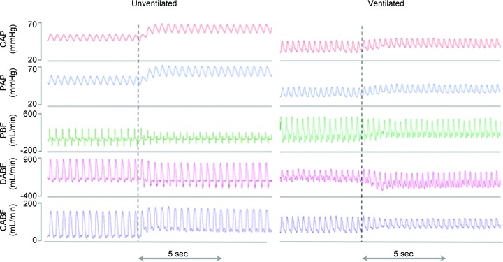
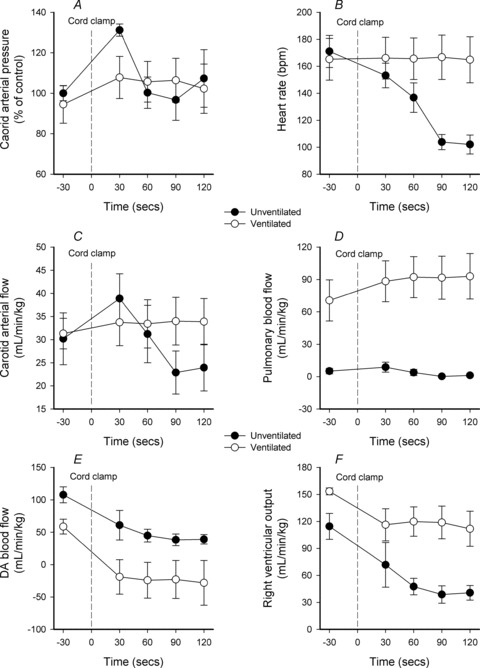
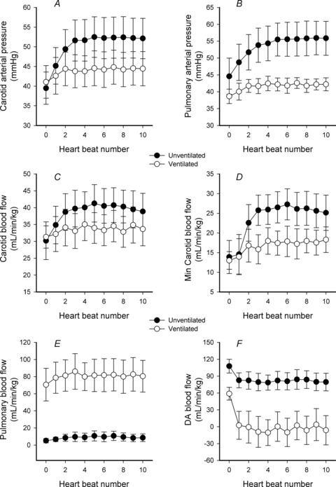
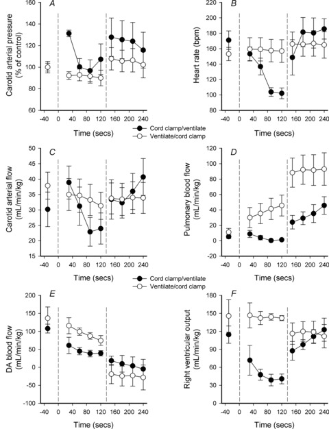
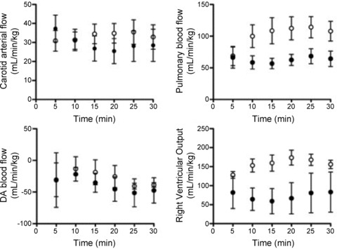
Delayed cord clamping improves circulatory stability in preterm infants at birth, but the underlying physiology is unclear. We investigated the effects of umbilical cord clamping, before and after ventilation onset, on cardiovascular function at birth. Prenatal surgery was performed on lambs (123 days) to implant catheters into the pulmonary and carotid arteries and probes to measure pulmonary (PBF), carotid (CaBF) and ductus arteriosus blood flows. Lambs were delivered at 126 ± 1 days and: (1) the umbilical cord was clamped at delivery and ventilation was delayed for about 2 min (Clamp 1st; n = 6), and (2) umbilical cord clamping was delayed for 3-4 min, until after ventilation was established (Vent 1st; n = 6). All lambs were subsequently ventilated for 30 min. In Clamp 1st lambs, cord clamping rapidly (within four heartbeats), but transiently, increased pulmonary and carotid arterial pressures (by ∼30%) and CaBF (from 30.2 ± 5.6 to 40.1 ± 4.6 ml min(-1) kg(-1)), which then decreased again within 30-60 s. Following ventilation onset, these parameters rapidly increased again. In Clamp 1st lambs, cord clamping reduced heart rate (by ∼40%) and right ventricular output (RVO; from 114.6 ± 14.4 to 38.8 ± 9.7 ml min(-1) kg(-1)), which were restored by ventilation. In Vent 1st lambs, cord clamping reduced RVO from 153.5 ± 3.8 to 119.2 ± 10.6 ml min(-1) kg(-1), did not affect heart rates and resulted in stable blood flows and pressures during transition. Delaying cord clamping for 3-4 min until after ventilation is established improves cardiovascular function by increasing pulmonary blood flow before the cord is clamped. As a result, cardiac output remains stable, leading to a smoother cardiovascular transition throughout the early newborn period.
Figures

Comment in
-
The art of cord clamping: sparing the linen or sparing the child?J Physiol. 2013 Apr 15;591(8):2021-2. doi: 10.1113/jphysiol.2013.253336. J Physiol. 2013. PMID: 23588499 Free PMC article. No abstract available.
References
-
- Alcorn DG, Adamson TM, Maloney JE, Robinson PM. A morphologic and morphometric analysis of fetal lung development in the sheep. Anat Rec. 1981;201:655–667. - PubMed
-
- Baenziger O, Stolkin F, Keel M, von Siebenthal K, Fauchere JC, Das Kundu S, Dietz V, Bucher HU, Wolf M. The influence of the timing of cord clamping on postnatal cerebral oxygenation in preterm neonates: a randomized, controlled trial. Pediatrics. 2007;119:455–459. - PubMed
-
- Chaparro CM, Lutter C. Beyond survival: integrated delivery care practices for longterm maternal and infant nutrition, health and development. Washington, DC: Pan American Health Organization; 2007.
-
- Dawes GS. Foetal and neonatal physiology: a comparative study of the changes at birth. Chicago: Year Book Medical Publishers, Inc; 1968.
Publication types
MeSH terms
LinkOut - more resources
Full Text Sources
Other Literature Sources
Medical
Research Materials
Miscellaneous