. 2013:19:319-32.
Epub 2013 Feb 7.

Cyclin-dependent kinase 5 promotes the stability of corneal epithelial cell junctions

Affiliations

Cyclin-dependent kinase 5 promotes the stability of corneal epithelial cell junctions

Parthasarathy Arpitha et al. Mol Vis. 2013.

Abstract

Purpose: Although cyclin-dependent kinase 5 (Cdk5) inhibits the formation of junctions containing N-cadherin, the effect of Cdk5 on junctions containing E-cadherin is less clear. The present study investigates the functional significance of Cdk5 in forming and maintaining cell-cell stability in corneal epithelial cells.

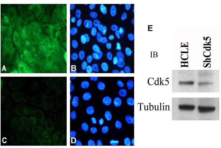
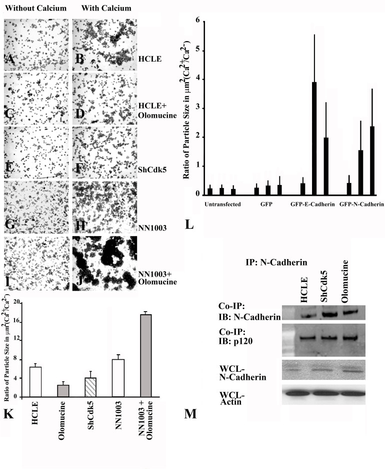
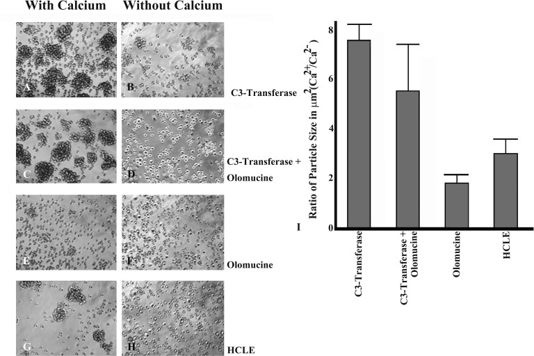
Methods: A Cdk5-deficient human corneal limbal epithelial cell line was generated by lentiviral transduction of small hairpin RNA specific for Cdk5 (shCdk5-HCLE cells). A blasticidin-inducible vector for expression of Cdk5-specific short hairpin RNA (ShCdk5) was generated by recombination and packaged into non-replicative lentiviral particles for transduction of human corneal limbal epithelial (HCLE) cells. Blasticidin-resistant cells were isolated for analysis. Cell aggregations were performed using HCLE, Cdk5 inhibitor olomoucine, ShCdk5, and MDA-MB 231 cells in the presence and absence of calcium, and particle size was measured using image analysis software. Relative protein concentrations were measured with immunoblotting and quantitative densitometry. Total internal reflection fluorescence (TIRF) microscopy was performed on cells transfected with green fluorescent protein (GFP)-E-cadherin or GFP-p120, and internalization of boundary-localized proteins was analyzed with particle tracking software. The stability of surface-exposed proteins was determined by measuring the recovery of biotin-labeled proteins with affinity chromatography. Rho and Rac activity was measured with affinity chromatography and immunoblotting.

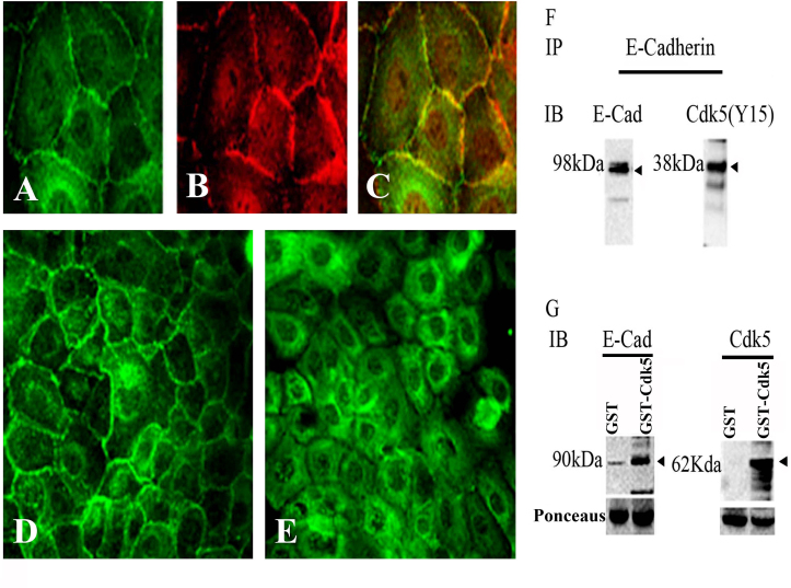
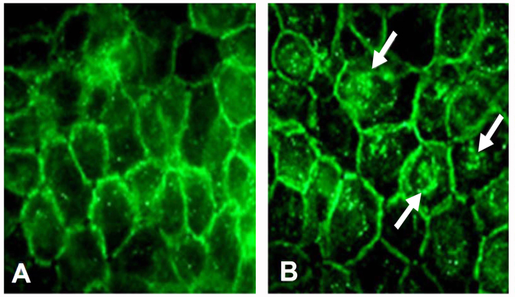
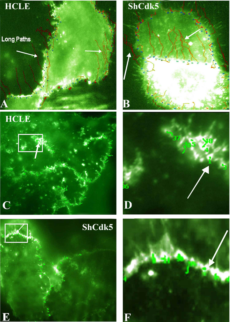
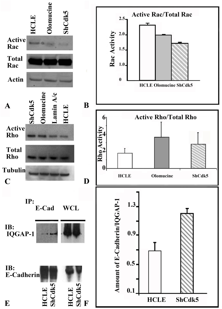
Results: Examining the effect of Cdk5 on E-cadherin containing epithelial cell-cell adhesions using a corneal epithelial cell line (HCLE), we found that Cdk5 and Cdk5 (pY15) coimmunoprecipitate with E-cadherin and Cdk5 (pY15) colocalizes with E-cadherin at cell-cell junctions. Inhibiting Cdk5 activity in HCLE or suppressing Cdk5 expression in a stable HCLE-derived cell line (ShHCLE) decreased calcium-dependent cell adhesion, promoted the cytoplasmic localization of E-cadherin, and accelerated the loss of surface-biotinylated E-cadherin. TIRF microscopy of GFP-E-cadherin in transfected HCLE cells showed an actively internalized sub-population of E-cadherin, which was not bound to p120 as it was trafficked away from the cell-cell boundary. This population increased in the absence of Cdk5 activity, suggesting that Cdk5 inhibition promotes dissociation of p120/E-cadherin junctional complexes. These effects of Cdk5 inhibition or suppression were accompanied by decreased Rac activity, increased Rho activity, and enhanced binding of E-cadherin to the Rac effector Ras GTPase-activating-like protein (IQGAP1). Cdk5 inhibition also reduced adhesion in a cadherin-deficient cell line (MDA-MB-231) expressing exogenous E-cadherin, although Cdk5 inhibition promoted adhesion when these cells were transfected with N-cadherin, as previous studies of Cdk5 and N-cadherin predicted. Moreover, Cdk5 inhibition induced N-cadherin expression and formation of N-cadherin/p120 complexes in HCLE cells.

Conclusions: These results indicate that loss of Cdk5 activity destabilizes junctional complexes containing E-cadherin, leading to internalization of E-cadherin and upregulation of N-cadherin. Thus, Cdk5 activity promotes stability of E-cadherin-based cell-cell junctions and inhibits the E-cadherin-to-N-cadherin switch typical of epithelial-mesenchymal transitions.


Figures

Figure 1
Figure 1
Co-localization of E-cadherin and cyclin dependent kinase5 (Cdk5) in the cell–cell borders. Human corneal limbal epithelial (HCLE) cells were grown on chamber slides, fixed, and immunostained stained with pY15 (green; A) and E-cadherin (red; B). Cyclin-dependent kinase 5 (Cdk5) and E-cadherin were localized to the cell–cell boundaries, and the overlay image demonstrates the colocalization (C) of E-cadherin and phosphorylated (pY15) CDK5. Confluent cultures of HCLE showing CDK5 border localization (D) confluent HCLE cultures when treated with olomoucine show a shift in the localization of pY15 CDK5 from the cell borders to the interior (E). E-cadherin and CDK5 form a part of the same protein complex (F, G) E-cadherin coimmuneprecipitates with CDK5 (38 kDa; F). Affinity chromatography for HCLE cell lysates were pulled down for glutathione-S-transferase-cyclin-dependent kinase 5 (GST-CDK5) and that glutathione beads bound to CDK5 formed a complex with E-cadherin (G). (A, B, C) 100X images and (D, E) 40X images.
Figure 2
Figure 2
Human corneal limbal epithelial cell line suppressed for cyclin-dependent kinase 5 expression. Immunofluorescence image showing staining for cyclin-dependent kinase 5 (CDK5) (green; A) in human corneal limbal epithelial (HCLE) cells and note the absence of Cdk5 in the ShCDK5 cells (C). Nuclear counterstaining 4', 6-diamidino-2-phenylindole (blue) for images in A and C (B, D). Lysates from these cells showing significant reduction in the expression levels of CDK5 in the plentiviral transduced HCLE (ShCDK5) cells (E). 100X images.
Figure 3
Figure 3
Cell–cell adhesions in the ShCDK5 cells. Confluent human corneal limbal epithelial (HCLE) and ShCDK5 cells immunostained with E-cadherin. The HCLE cells show border localization of E-cadherin (A). ShCDK5 increases the border localization to the cell interior marked by punctuate staining (arrows) within the cells. The ratio of the border to cell interior was significantly reduced in the ShCDK5 E-cadherin-labeled cells (0.347±0.145) when compared to the HCLE (0.535±0.142) 100X images.
Figure 4
Figure 4
Cell-cell junction formation in the ShCDK5 human corneal limbal epithelial cells. Human corneal limbal epithelial (HCLE) cells (A, B), in the absence of cyclin-dependent kinase 5 (CDK5) with olomoucine (C, D) or ShCDK5 (E, F) and lens epithelial cells (NN1003; G, H) were treated with olomoucine (I, J). The cells were allowed to form cell–cell adhesions in the presence or absence of calcium for 1 h at 37 °C, and particle aggregates were cytospin on to glass slides to measure the size of the aggregates. Calcium-dependent E-cadherin junctions were formed in the presence of CDK5 (B, K) in the HCLE cells in contrast to N-cadherin expressing lens epithelial cells that form larger aggregates in the absence of CDK5 (J, K). (AJ) 20X images. E-cadherin and N-cadherin induction during cell–cell junction formation in the MDA-MB-231 cells (L). Green fluorescent protein (GFP)-E-cadherin and GFP-N-cadherin transfected cells were allowed to form cell–cell aggregates in the absence of calcium (lane 1), presence (lane 2) and with olomoucine (lane 3). Cells containing E-cadherin form more cell–cell aggregates (group 3) while the N-cadherin (group 4) aggregates increases in the presence of olomoucine. N-cadherin induction in the ShCDK5 and olomoucine HCLE (M) CDK5 inhibition leading to degradation of E-cadherin leads to induction of N-cadherin. P120 coimmuneprecipitates with N-cadherin to stabilize the junctional complex in the ShCDK5 and olomoucine-treated HCLE. IP=immunoprecipitation; WCl=whole cell lysate.
Figure 5
Figure 5
Total internal reflection fluorescence analysis for the E-cadherin internalization pathway in the human corneal limbal epithelial and ShCDK5 cells. Green fluorescent protein (GFP)-E-cadherin transfected (AB) epithelial cells with and without olomoucine and ShCDK5 cultures were analyzed for E-cadherin particle movement for distance and tortuosity from the cell–cell borders at a constant time (A). The tortuosity or particle tracking of the E-cadherin-containing vesicles was recorded. The E-cadherin vesicles moved in long paths (arrows) from the cell borders to the interior, and such long paths were significantly greater in the ShCDK5 suggestive of internalization than the control human corneal limbal epithelial (HCLE) cells. Total internal reflection fluorescence (TIRF) analysis for the p120 (CH) shows internalization pathway in the HCLE and ShCDK5. A GFP-p120 clone was transfected into the HCLE and ShCDK5 cells, and the vesicle tracking (arrows) for the tortuosity was measured. P120 vesicles remained in the cell–cell borders, and no long paths were internalized. In the HCLE and ShCDK5 cells, the p120 tortuosity paths were restricted within the cell–cell borders. Panel inserts in the HCLE cells transfected with p120 (C) and ShCdk5 transfected with p120 (E) are enlarged (zoom to a factor of 2) and shown in D and F, respectively, along with the tortuosity path for each junctional vesicle analyzed. The distance and tortuosity of paths by the E-cadherin- and p120-containing particles (n>100) were tracked at a constant time for all the particles analyzed using TIRF microscopy are described in Table 1. The E-cadherin-containing particles in the ShCdk5 cells moved fast, and these long, straight paths were internalized (B). The p120-containing particles in the ShCdk5 cells moved slowly and stayed near the cell–cell boundary (C–F). In the absence of CDK5, the E-cadherin and p120 junctional complex dissociate, promoting internalization and endocytosis of E-cadherin. Cdk5 stabilizes the junction by preventing E-cadherin-containing vesicles from being endocytosed. 100X images.
Figure 6
Figure 6
Rac activity is required for cell–cell adhesions in the human corneal limbal epithelial cells. The ShCDK5 human corneal limbal epithelial (HCLE) and olomoucine-treated cultures reduced Rac activity (A) leading to instability of the cell–cell adhesion junctions, while the HCLE cells have significantly higher Rac activity (B) Rho kinase activity was tested in the in ShCDK5 HCLE cells. C: Representative experiment showing a twofold increase in the Rho activity in confluent cultures of ShCDK5 and in olomoucine-treated cells when compared to the control Lamin A/C and HCLE. Significant (n=3; p≤0.05) increase in the Rho activity in the absence of Cdk5 with olomoucine (D) and decrease in Rac activity in the ShCdk5 and olomoucine, suggesting destabilization of cell–cell adhesions in the olomoucine and ShCDK5 cells, is represented in the graph (B, D). A cytoskeletal Rac modulator, IQGAP1, coimmunes precipitates with E-cadherin (E, F) in the ShCDK5 suggesting the internalization and degradation of E-cadherin. Stable cell–cell adhesions in HCLE cells are marked by significant reduction in the interaction of IQGAP1 with E-cadherin. (E, F). Note: Rho or Rac activity was measured as individual values normalized with the tubulin/actin and ratio of active Rho or active Rac against total Rho or total Rac, respectively. IB=immunoblot; IP=immunoprecipitation.
Figure 7
Figure 7
Rho activity mediates the downstream regulation of CDK5 in promoting cell–cell junction formation. Cell aggregates were measured with and without Ca2+ and defined as particle size in μm2 using NIH image J. In the presence of the C3 transferase, a Rho kinase inhibitor, the human corneal limbal epithelial (HCLE) cells form larger cell aggregates (A), and there is a reduction in the particle size in the presence of CDK5 and Rho kinase inhibitors suggesting the role of Cdk5 downstream signaling via the Rho kinase in the cell–cell junction formation (C). Olomoucine alone (E) reduces the formation of cell–cell aggregates than the control untreated (G) HCLE. Ratio of particle size=particle size (um) in the presence of calcium/ particle size (um) without calcium is represented in the graph (I). AH are 20X images.

Similar articles

Cited by

References

    1. Meyerson G, Pfenninger KH, Påhlman S. A complex consisting of pp60c-src/pp60c-srcN and a 38 kDa protein is highly enriched in growth cones from differentiated SH-SY5Y neuroblastoma cells. J Cell Sci. 1992;103:233–43. - PubMed
    1. Gao CY, Stepp MA, Fariss R, Zelenka P. Cdk5 regulates activation and localization of Src during corneal epithelial wound closure. J Cell Sci. 2004;117:4089–98. - PubMed
    1. Rosales JL, Lee KY. Extraneuronal roles of cyclin-dependent kinase 5. Bioessays. 2006;28:1023–34. Review. - PubMed
    1. Tsai LH, Delalle I, Caviness VS, Jr, Chae T, Harlow E. p35 is a neural-specific regulatory subunit of cyclin-dependent kinase 5. Nature. 1994;371:419–23. - PubMed
    1. Humbert S, Dhavan R, Tsai L. p39 activates cdk5 in neurons, and is associated with the actin cytoskeleton. J Cell Sci. 2000;113:975–83. - PubMed

Publication types

MeSH terms

LinkOut - more resources