An endothelialized urothelial cell-seeded tubular graft for urethral replacement
- PMID: 23401738
- PMCID: PMC3559612
- DOI: 10.5489/cuaj.12217
An endothelialized urothelial cell-seeded tubular graft for urethral replacement
Abstract
Introduction: Many efforts are used to improve surgical techniques and graft materials for urethral reconstruction. We developed an endothelialized tubular structure for urethral reconstruction.
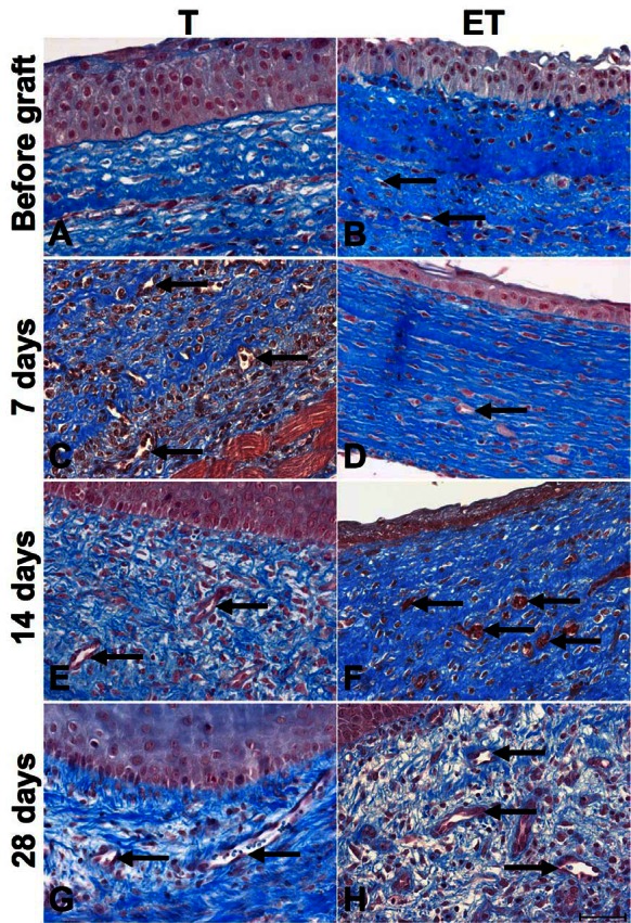
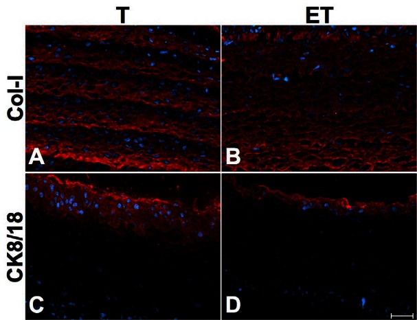
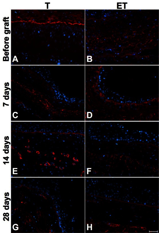
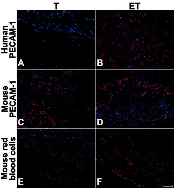
Methods: Two tubular models were created in vitro. Human fibroblasts were cultured for 4 weeks to form fibroblast sheets. Then, endothelial cells (ECs) were seeded on the fibroblast sheets and wrapped around a tubular support to form a cylinder for the endothelialized tubular urethral model (ET). No ECs were added in the standard tubular model (T). After 21 days of maturation, urothelial cells were seeded into the lumen of both models. Constructs were placed under perfusion in a bioreactor for 1 week. At several times, histology and immunohistochemistry were performed on grafted nude mice to evaluate the impact of ECs on vascularization.
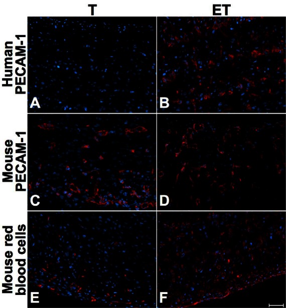
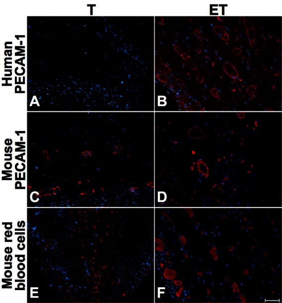
Results: Both models produced an extracellular matrix, without exogenous material, and developed a pseudostratified urothelium. Seven days after the graft, mouse red blood cells were present only in the outer layers in T model, but in the full thickness of ET model. After 14 days, erythrocytes were present in both models, but in a greater proportion in ET model. At day 28, both models were well-vascularized, with capillary-like structures in the whole thickness of the tubes.
Conclusion: Incorporating endothelial cells was associated with an earlier vascularization of the grafts, which could decrease the necrosis of the transplanted tissue. As those models can be elaborated with the patient's cells, this tubular urethral graft would be unique in its autologous property.
Figures


Similar articles
-
Surgical option for the correction of Peyronie's disease: an autologous tissue-engineered endothelialized graft.J Sex Med. 2011 Nov;8(11):3227-35. doi: 10.1111/j.1743-6109.2011.02374.x. Epub 2011 Jun 30. J Sex Med. 2011. PMID: 21718447
-
Tissue engineering of a genitourinary tubular tissue graft resistant to suturing and high internal pressures.Tissue Eng Part A. 2009 Jan;15(1):197-202. doi: 10.1089/ten.tea.2007.0303. Tissue Eng Part A. 2009. PMID: 18759664
-
Gel Casting as an Approach for Tissue Engineering of Multilayered Tubular Structures.Tissue Eng Part C Methods. 2020 Mar;26(3):190-198. doi: 10.1089/ten.TEC.2019.0280. Tissue Eng Part C Methods. 2020. PMID: 32089096
-
The role of vascularization in nerve regeneration of nerve graft.Neural Regen Res. 2020 Sep;15(9):1573-1579. doi: 10.4103/1673-5374.276327. Neural Regen Res. 2020. PMID: 32209756 Free PMC article. Review.
-
A method for constructing vascularized muscle flap.Methods. 2015 Aug;84:70-5. doi: 10.1016/j.ymeth.2015.03.021. Epub 2015 Apr 2. Methods. 2015. PMID: 25843607 Review.
Cited by
-
Engineered human organ-specific urethra as a functional substitute.Sci Rep. 2022 Dec 9;12(1):21346. doi: 10.1038/s41598-022-25311-1. Sci Rep. 2022. PMID: 36494468 Free PMC article.
-
Biological Assessment of Zn-Based Absorbable Metals for Ureteral Stent Applications.Materials (Basel). 2019 Oct 12;12(20):3325. doi: 10.3390/ma12203325. Materials (Basel). 2019. PMID: 31614757 Free PMC article.
-
Human Organ-Specific 3D Cancer Models Produced by the Stromal Self-Assembly Method of Tissue Engineering for the Study of Solid Tumors.Biomed Res Int. 2020 Apr 13;2020:6051210. doi: 10.1155/2020/6051210. eCollection 2020. Biomed Res Int. 2020. PMID: 32352002 Free PMC article. Review.
-
Genitourinary Tissue Engineering: Reconstruction and Research Models.Bioengineering (Basel). 2021 Jul 13;8(7):99. doi: 10.3390/bioengineering8070099. Bioengineering (Basel). 2021. PMID: 34356206 Free PMC article. Review.
-
Tissue Engineering for Gastrointestinal and Genitourinary Tracts.Int J Mol Sci. 2022 Dec 20;24(1):9. doi: 10.3390/ijms24010009. Int J Mol Sci. 2022. PMID: 36613452 Free PMC article. Review.
References
LinkOut - more resources
Full Text Sources
Research Materials