. 2013 Apr;33(8):1594-607.
doi: 10.1128/MCB.01220-12. Epub 2013 Feb 11.

P19ARF and RasV¹² offer opposing regulation of DHX33 translation to dictate tumor cell fate

Affiliations

P19ARF and RasV¹² offer opposing regulation of DHX33 translation to dictate tumor cell fate

Yandong Zhang et al. Mol Cell Biol. 2013 Apr.

Abstract

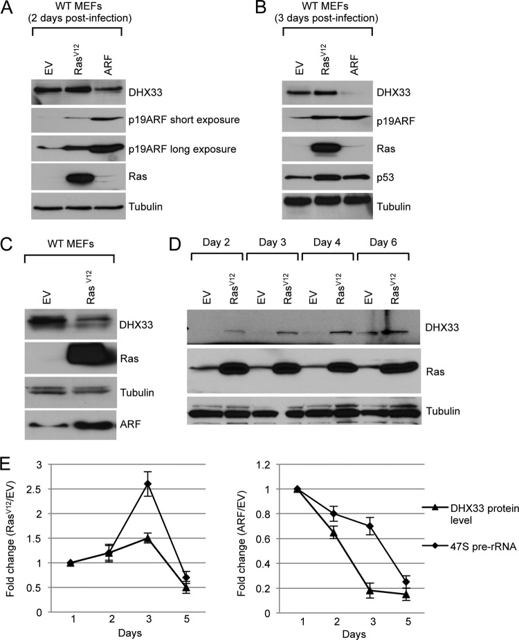
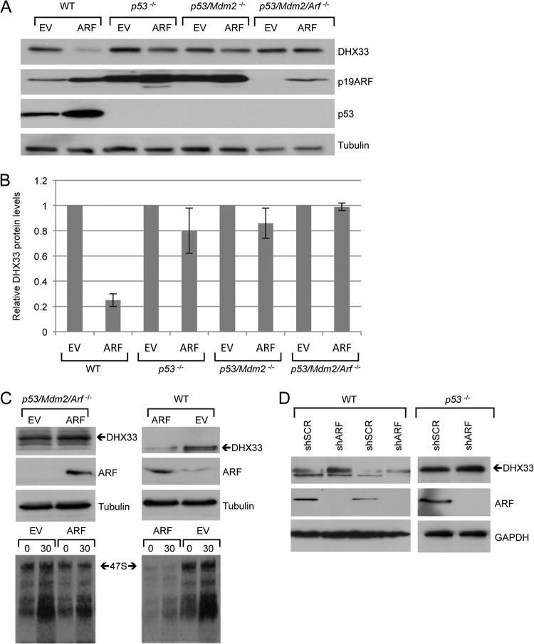
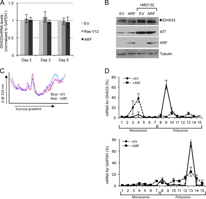
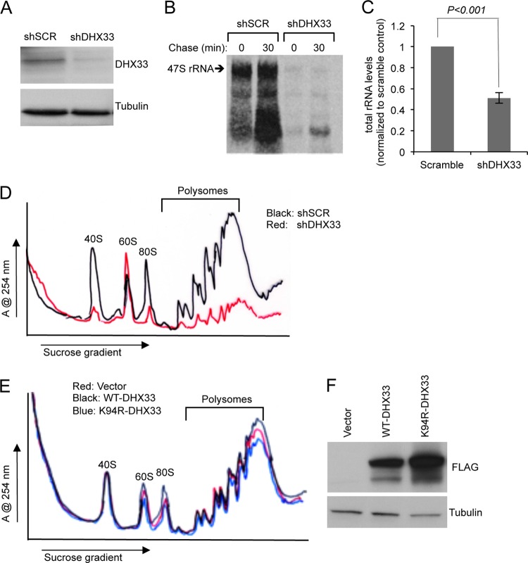
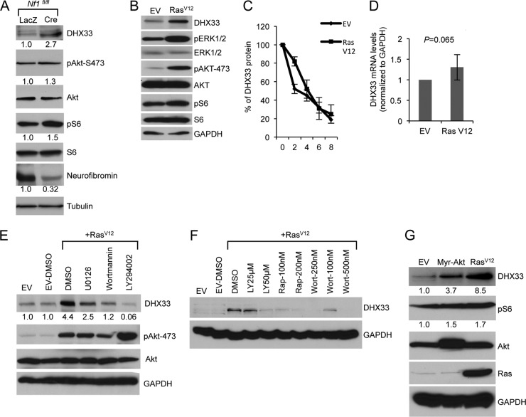
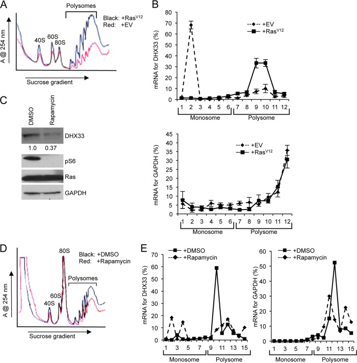
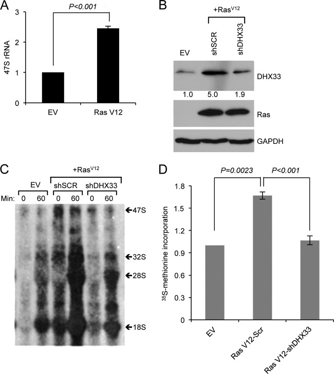
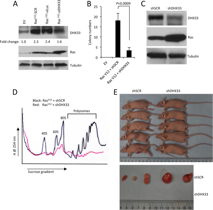
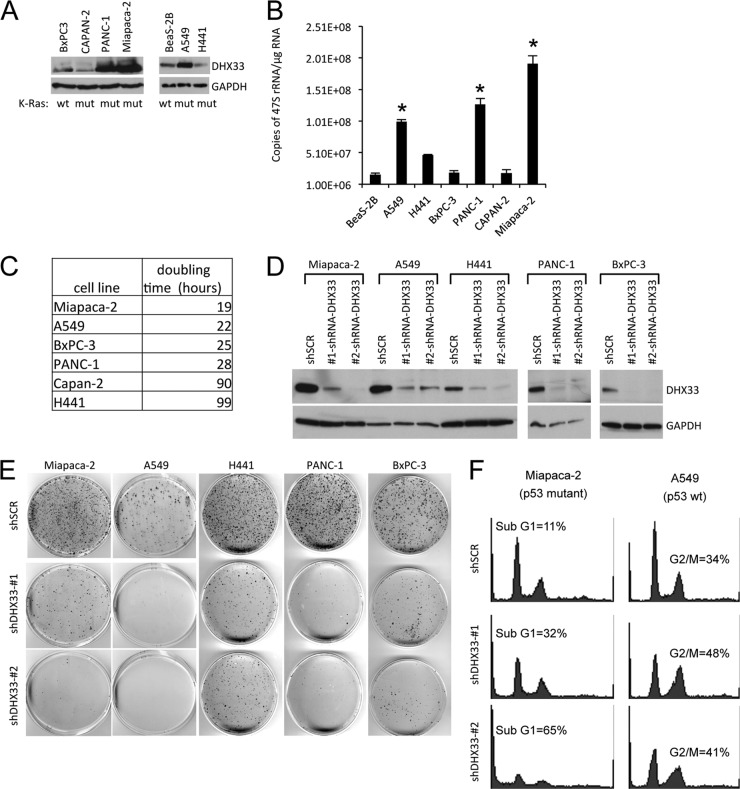
DHX33 is a pivotal DEAH-box RNA helicase in the multistep process of RNA polymerase I-directed transcription of the ribosomal DNA locus. We explored the regulation of DHX33 expression by Ras(V12) and ARF to determine DHX33's role in sensing these opposing signals to regulate ribosome biogenesis. In wild-type primary fibroblasts, Ras(V12) infection induced a transient increase in DHX33 protein level, as well as an rRNA transcriptional rate that was eventually suppressed by a delayed activation of the ARF/p53 pathway. DHX33 expression was exclusively controlled at the level of translation. ARF caused a dramatic reduction in polysome-associated DHX33 mRNAs, while Ras(V12) led to a complete shift of existing DHX33 mRNAs to actively translating polysomes. The translation of DHX33 by Ras(V12) was sensitive to inhibitors of phosphatidylinositol 3-kinase, mTOR, and mitogen-activated protein and was pivotal for enhanced rRNA transcription and enhanced overall cellular protein translation. In addition, DHX33 knockdown abolished Ras(V12)-induced rRNA transcription and protein translation and prevented both the in vitro and in vivo transforming properties of oncogenic Ras(V12). Our results directly implicate DHX33 as a crucial player in establishing rRNA synthesis rates in the face of Ras(V12) or ARF signals, adjusting ribosome biogenesis to match the appropriate growth or antigrowth signals.


Figures

Fig 1
Fig 1
Regulation of DHX33 during oncogenic stress. (A) Wild-type MEFs were infected with retroviruses encoding RasV12, p19ARF, or empty vector, whole-cell lysates were prepared at 2 days postinfection and were subjected to Western blot analysis with the indicated antibodies. (B) The above-mentioned cells were harvested 3 days postinfection, and whole-cell extracts were subjected to Western blot analysis with the indicated antibodies. (C) Wild-type MEFs were infected with retroviruses encoding RasV12 or empty vector, whole-cell extracts were prepared 5 days postinfection and were subjected to Western blot analysis with the indicated antibodies. (D) Arf-null MEFs were infected with retroviruses encoding RasV12 or empty vector, whole-cell extracts were prepared from 2 days till 6 days postinfection and were subjected to Western blot analysis with the indicated antibodies. (E) Wild-type MEFs were infected with the above-mentioned retroviruses, and total RNA was extracted at 2, 3, or 5 days postinfection. Mouse 47S pre-rRNA levels were analyzed by real-time PCR and graphed in a time-dependent manner. Changes in DHX33 protein levels in the time course were also graphed after quantitation of DHX33 signals in panels A to C after normalization to the empty vector control. Error bars were taken from three independent experiments.
Fig 2
Fig 2
Reduction of DHX33 by ARF infection is dependent on p53 and Mdm2. (A) Wild-type MEFs, p53-null MEFs, p53−/−; Mdm2−/− MEFs, and p53−/−; Mdm2−/−; Arf−/− MEFs were infected with retroviruses encoding either pBABE empty vector or pBABE-HA-ARF. Whole-cell lysates were prepared 5 days postinfection and were subjected to Western blot analysis by the indicated antibodies. (B) Quantitation of the DHX33 protein levels after normalization to empty vector in each group, error bars were taken from three independent experiments. (C) MEFs were infected by retrovirus encoding pBABE empty vector or pBABE-HA-ARF. At 4 days postinfection, cells were harvested and analyzed by Western blotting with the indicated antibodies. At 4 days postinfection, cells were also pulsed by [3H]uridine and chased at the indicated time points. Total RNA were analyzed for 47S pre-rRNA levels. (D) WT MEFs or p53-null MEFs were infected with lentivirus encoding either shSCR or shARF. At 4 days postinfection, the cells were harvested and subjected to Western blot analysis with the indicated antibodies.
Fig 3
Fig 3
Induction of ARF inhibits DHX33 translation. (A) Wild-type MEFs were infected with retroviruses encoding empty vector, p19ARF, or RasV12, and the total RNA was extracted from each sample at 2, 3, or 5 days postinfection. DHX33 mRNA levels were analyzed by qPCR with GAPDH as an internal control. Bars represent the standard deviation taken from three separate experiments. (B) Wild-type MEFs were infected with retroviruses encoding empty vector (EV) or p19ARF. At 3 days postinfection, the cells were treated with 50 μM MG132 for 6 h, and total cell lysates were prepared and subjected to Western blot analysis with the indicated antibodies. p21 protein stabilization was used as a positive control to monitor MG132 function. (C) A total of 1.5 × 10⁶ wild-type cells infected with retroviruses encoding empty vector or p19ARF at 3 days postinfection were subjected to cytosolic polysome fractionation. The absorbance was monitored at 254 nm, and resultant ribosome profiles are shown for each sample. (D) The above-mentioned fractions from monoribosomes or polysomes were subjected to total RNA isolation and qPCR analysis to detect DHX33 mRNA levels. GAPDH mRNA levels were used as a control. The data presented are the percentages of mRNA from each fraction calculated from a standard curve generated by a series of diluted DHX33 plasmid. Error bars were taken from two independent experiments.
Fig 4
Fig 4
DHX33 protein knockdown or overexpression influences ribosome biogenesis and protein translation. (A) Arf/p53/Mdm2−/− MEFs were infected by lentivirus encoding shRNA-DHX33 or shScrambled (shSCR). At 3 days after infection, the cell lysates were subjected to Western blot analysis with anti-DHX33 and tubulin antibodies. (B) Infected cells from above were pulsed with [3H]uridine and chased for the indicated time points to monitor newly synthesized rRNA. Equal numbers of cells were pelleted for total RNA extraction. RNA was separated and transferred onto nylon membranes for autoradiography. (C) Equal numbers of the above-mentioned cells were subjected to total RNA isolation and then isolated by formaldehyde RNA denaturing gel. 28S and 18S rRNA were visualized by ethidium bromide staining and quantified. Bars were taken from three different experiments. (D) Equal numbers of Arf/p53/Mdm2−/− MEFs infected by the indicated short-hairpin lentiviruses were subjected to cytosolic ribosome profile analysis at 4 days postinfection. (E and F) Arf/p53/Mdm2−/− MEFs were infected with lentiviruses encoding empty vector, DHX33 (wild type) or mutant DHX33 (K94R). At 4 days postinfection, infected cells were subjected to cytosolic polysome profile analysis (E) and Western blot analysis with the indicated antibodies (F).
Fig 5
Fig 5
Ras activity induces DHX33 protein expression. (A) Nf1fl/fl MEFs were infected with adenoviruses encoding either LacZ or Cre recombinase at a multiplicity of infection of 200. At 2 days postinfection, the cells were then serum starved for 72 h. Equal amount of cell lysates were subjected to Western blot analysis with the indicated antibodies. (B) Arf-null ear fibroblasts from 2-month-old mice were infected with retroviruses encoding either pBABE empty vector (EV) or pBABE-RasV12. At 3 days postinfection, infected cells were subjected to Western blot analysis with the indicated antibodies. (C) The above-mentioned cells were treated with cycloheximide at a concentration of 80 μg/ml for the indicated times. Protein extracts from the cells pelleted from the indicated time points was subjected to Western blot analysis. Signals of DHX33 protein was graphed after normalization to GAPDH control. Bars represent errors from two independent experiments. (D) Total RNA was isolated from the above-mentioned cells and changes of DHX33 mRNA levels were analyzed by qPCR with GAPDH as a control. P is derived from five separate experiments. (E) Arf-null ear fibroblasts were infected with empty vector or RasV12. At 3 days postinfection, the cells were treated with U0126 (20 μM), wortmannin (100 nM), or LY294002 (50 μM) for 24 h. Cell lysates were subjected to Western blot analysis with the indicated antibodies. (F) Arf-null cells infected with empty vector or RasV12 were treated with rapamycin, wortmannin, or LY294002 as indicated for 24 h. Cell lysates were prepared and analyzed for DHX33 protein levels with GAPDH as a loading control. (G) Arf-null MEFs were infected with retroviruses encoding myristoylated Akt (Myr-Akt), RasV12, or empty vector. Cell lysates were prepared at 4 days postinfection after puromycin selection and analyzed by Western blotting for DHX33, pAkt-473, Akt, and GAPDH protein levels. The fold change is indicated below identified blots.
Fig 6
Fig 6
DHX33 protein induction is under translational control. (A) A total of 3 × 10⁶ Arf-null cells infected with retroviruses encoding empty vector or RasV12 at 4 days postinfection were subjected to cytosolic ribosome profiling. (B) The resultant fractionations from above were analyzed by RT-PCR for DHX33 mRNA distribution on ribosomes. GAPDH was used as a negative control. Bar data were taken from three independent experiments. (C) RasV12-infected Arf-null cells at 4 days postinfection were treated with rapamycin at 100 nM for 24 h. Whole-cell extracts were then subjected to total protein analysis by Western blotting with the indicated antibodies. The fold change is indicated underneath the blots. (D) A total of 3 × 10⁶ of RasV12-infected Arf-null cells after rapamycin treatment (100 nM) were subjected to cytosolic ribosome profiling. (E) The resultant fractions from above were analyzed by RT-PCR for DHX33 mRNA distribution on ribosomes. GAPDH was used as a negative control. The data represents a typical result from three independent experiments.
Fig 7
Fig 7
DHX33 protein induction plays a crucial role in RasV12-enhanced rRNA transcription. (A) Arf-null ear fibroblasts from 2-month-old mice were infected with either empty vector or RasV12-encoding retroviruses. Total RNA was extracted and analyzed by RT-PCR for 47S pre-rRNA levels. Error bars indicate standard deviation from three independent experiments. (B, C, and D) Arf-null ear fibroblasts were infected with retroviruses encoding empty vector or RasV12. At 2 days postinfection, the cells were then infected with lentiviruses encoding shScrambled (SCR) or shRNA-DHX33 for 3 days. Cells were then subjected to Western blot analysis for DHX33 protein levels (B). Equal numbers of cells were pulsed with [3H]uridine and chased at the indicated time points, the total RNA was isolated and separated for rRNA synthesis analysis, and a representative result from three independent experiments is shown (C). The cells were then pulse-labeled with [35S]methionine incorporation and 35S-labeled proteins were measured. Error bars represent the standard deviation from three independent experiments (D). **, P < 0.001.
Fig 8
Fig 8
DHX33 induction is required for RasV12-initiated tumor formation. (A) Arf-null ear fibroblasts from 2-month-old mice were infected with retroviruses encoding pBABE-empty vector (EV) or pBABE-RasV12. Cells were then infected with lentiviruses encoding shScrambled (shSCR), shLuciferase (shLuc), or shDHX33. Whole-cell lysates were extracted and analyzed by Western blotting with Ras, DHX33, and tubulin antibodies. (B) A total of 5 × 10³ infected cells were plated onto soft agar 60-mm plates in triplicate to measure anchorage-independent cell growth after 14 days. Quantitation of the colony numbers is presented from three representative fields under ×4 magnification. Error bars represent the standard deviation calculated from three different fields of colonies on triplicate plates. (C) Arf-null NIH 3T3 cells were infected with retroviruses encoding RasV12, followed by infection with lentiviruses encoding shScrambled (shSCR) or shDHX33. Whole-cell lysates were subjected to Western blot analysis with Ras, DHX33, and tubulin antibodies. (D) A total of 3 × 10⁶ infected NIH 3T3 cells were subjected to cytosolic ribosome profiles. (E) The upper panel shows NIH 3T3 cells infected with retroviruses encoding RasV12 that were then infected with shSCR or shDHX33 lentiviruses. A total of 106 infected NIH 3T3 cells were injected into the flanks of nude mice. Tumor formation was visualized and photographed after 14 days. For the lower panel, mice were sacrificed at day 14 postinjection, and tumors were excised and photographed.
Fig 9
Fig 9
DHX33 is overexpressed in Ras-mutated cancer cell lines and is required for their efficient growth and proliferative properties. (A) A panel of K-Ras mutated or wild-type cancer cell lines (mutation status is shown at the bottom) were screened for total DHX33 protein expression, p14ARF status is also shown at the bottom. (B) 47S rRNA was measured by RT-PCR and normalized to total RNA levels. Error bars represent the standard deviation from three separate experiments. *, P < 0.001. (C) A total of 5 × 10⁴ cells were plated onto six-well culture plates. Cell numbers were counted daily and graphed. The doubling time was calculated based on growth curves and is shown in the table. (D) The indicated cell lines were infected with lentiviruses encoding shScrambled (shSCR) or shDHX33. Whole-cell lysates were extracted 4 days postinfection and subjected to Western blot analysis with antibodies recognizing DHX33 and tubulin. (E) shSCR or shDHX33-infected cells (104) from indicated cancer cell lines were plated onto 100-mm culture dishes. The cells were fixed 10 or 20 days later with 100% methanol and incubated with Giemsa stain for 1 h. Stained colonies were air dried and photographed. (F) shSCR or shDHX33-infected cells (104) from Miapaca-2 and A549 cancer cells were subjected to cell cycle analysis by flow cytometry after propidium iodide staining.

Similar articles

Cited by

References

    1. Hanahan D, Weinberg RA. Hallmarks of cancer: the next generation. Cell 144:646–674 - PubMed
    1. Luo J, Solimini NL, Elledge SJ. 2009. Principles of cancer therapy: oncogene and non-oncogene addiction. Cell 136:823–837 - PMC - PubMed
    1. Drygin D, Rice WG, Grummt I. The RNA polymerase I transcription machinery: an emerging target for the treatment of cancer. Annu. Rev. Pharmacol. Toxicol. 50:131–156 - PubMed
    1. Maggi LB, Jr, Weber JD. 2005. Nucleolar adaptation in human cancer. Cancer Invest. 23:599–608 - PMC - PubMed
    1. Montanaro L, Trere D, Derenzini M. 2008. Nucleolus, ribosomes, and cancer. Am. J. Pathol. 173:301–310 - PMC - PubMed

Publication types

MeSH terms

Substances