. 2013 Apr;33(8):1632-44.
doi: 10.1128/MCB.01503-12. Epub 2013 Feb 11.

The MCM8-MCM9 complex promotes RAD51 recruitment at DNA damage sites to facilitate homologous recombination

Affiliations

The MCM8-MCM9 complex promotes RAD51 recruitment at DNA damage sites to facilitate homologous recombination

Jonghoon Park et al. Mol Cell Biol. 2013 Apr.

Abstract

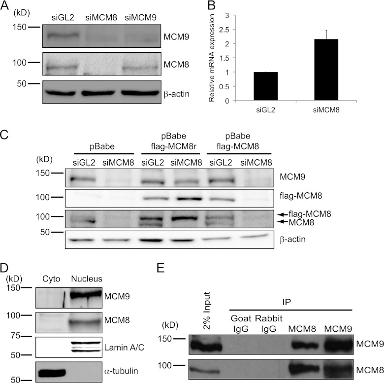
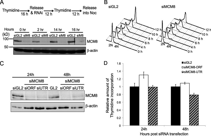
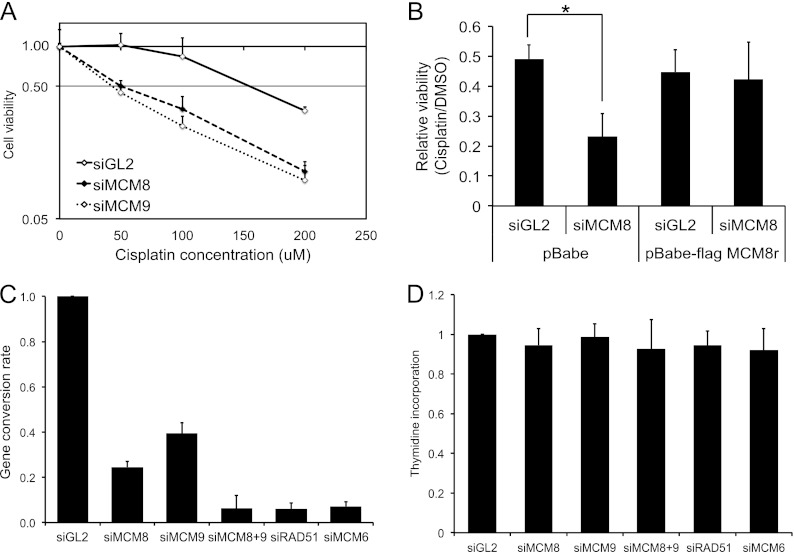
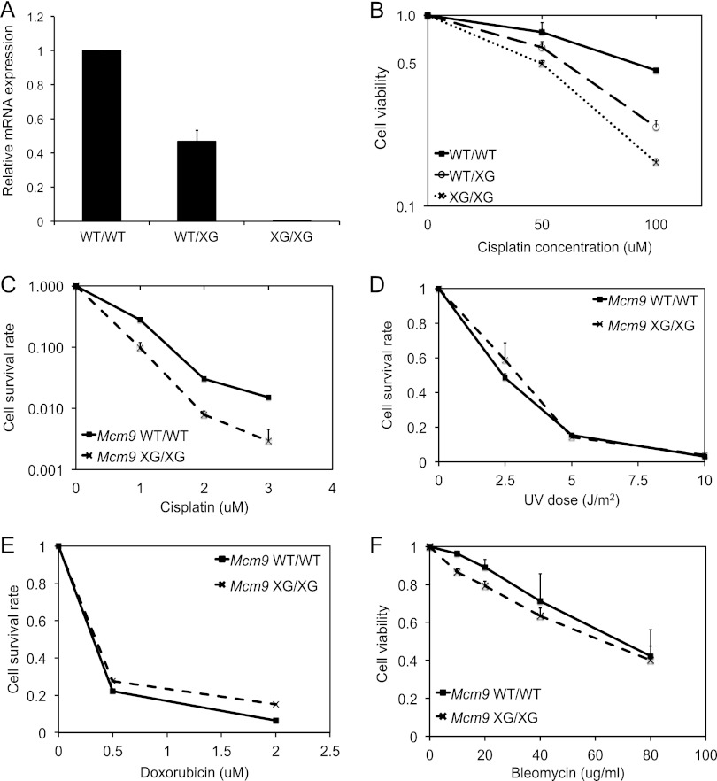
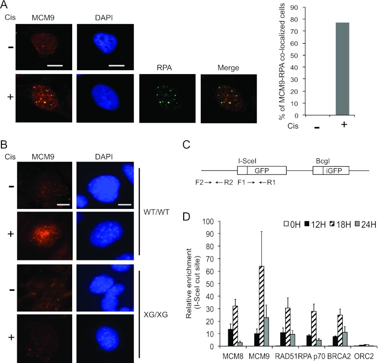
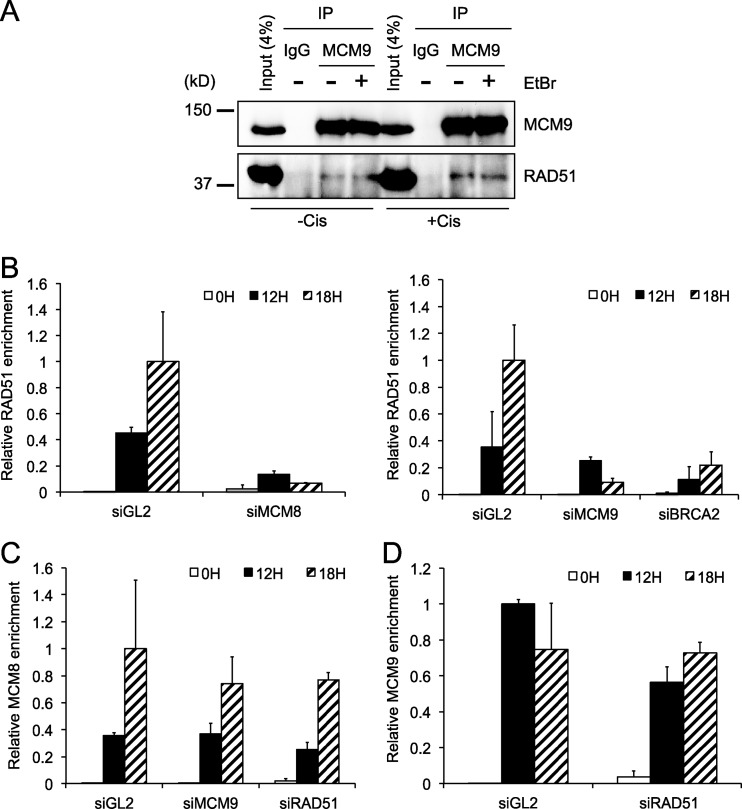
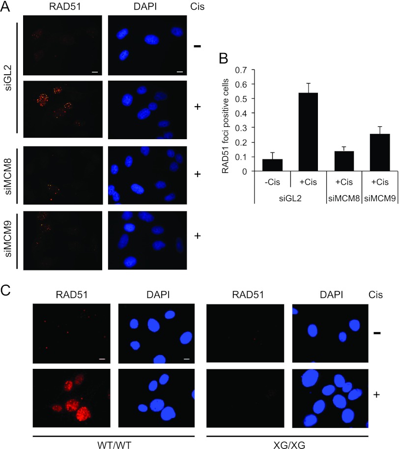
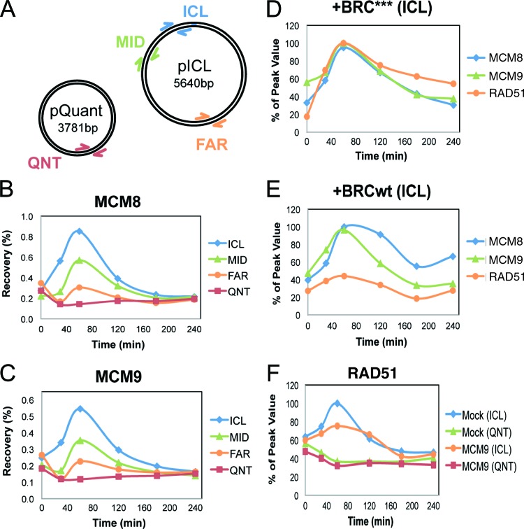
The minichromosome maintenance protein homologs MCM8 and MCM9 have previously been implicated in DNA replication elongation and prereplication complex (pre-RC) formation, respectively. We found that MCM8 and MCM9 physically associate with each other and that MCM8 is required for the stability of MCM9 protein in mammalian cells. Depletion of MCM8 or MCM9 in human cancer cells or the loss of function MCM9 mutation in mouse embryo fibroblasts sensitizes cells to the DNA interstrand cross-linking (ICL) agent cisplatin. Consistent with a role in the repair of ICLs by homologous recombination (HR), knockdown of MCM8 or MCM9 significantly reduces HR repair efficiency. Chromatin immunoprecipitation analysis using human DR-GFP cells or Xenopus egg extract demonstrated that MCM8 and MCM9 proteins are rapidly recruited to DNA damage sites and promote RAD51 recruitment. Thus, these two metazoan-specific MCM homologs are new components of HR and may represent novel targets for treating cancer in combination with DNA cross-linking agents.


Figures

Fig 1
Fig 1
MCM8 interacts with MCM9 and forms a stable nuclear complex. (A) Immunoblots of lysates of U2OS cells transfected with siRNA targeting the luciferase gene (siGL2, control) or the Mcm8 or Mcm9 genes (siMCM8 [siMCM8-ORF] and siMCM9, respectively). β-Actin protein serves as a loading control. (B) qRT-PCR of Mcm9 mRNA using total RNA from U2OS cells after siRNA transfection, normalized to GAPDH mRNA. Means + the standard deviations (SD) of triplicate measurements are shown. (C) Ectopic expression of siMCM8-resistant MCM8 protein (MCM8r) restores MCM9 levels in U2OS cells depleted of endogenous MCM8 protein. Wild-type and N-terminally Flag-tagged MCM8 (flag-MCM8) or siRNA resistant Flag-MCM8 protein (flag-MCM8r) were stably expressed in U2OS cells and transfected with siRNA targeting endogenous MCM8 or ectopic Flag-MCM8 but not ectopic MCM8r. Lysates were harvested 48 h after transfection and analyzed by SDS-PAGE and Western blotted with antibodies against the indicated proteins. The β-actin blot shows equal protein loading. (D) MCM8 and MCM9 are nuclear proteins. Immunoblots of cytoplasmic and nuclear proteins from 293T cells show that MCM8 and MCM9 are nuclear proteins. α-Tubulin and lamin A/C were used as controls of fractionation, respectively. Cyto, cytoplasmic proteins; Nucleus, nuclear proteins. (E) MCM8 associates with MCM9. Immunoprecipitates of endogenous MCM8 or MCM9 from 293T nuclear lysates coimmunoprecipitate MCM9 or MCM8, respectively.
Fig 2
Fig 2
MCM8-9 is not essential for DNA replication. (A) Schematic of the synchronization procedure used in U2OS cells. The extent of MCM8 protein knockdown by siRNA at each indicated time point following release from the double thymidine block was tested by SDS-PAGE and immunoblotting. β-Actin is shown as a loading control. siM8, siMCM8-UTR. (B) Propidium iodide fluorescence-activated cell sorting histograms are shown at the indicated hours after release from double thymidine block. (C) U2OS cells were harvested at the indicated time point after transfection of the indicated siRNAs. An immunoblot shows the amount of MCM8, and β-actin serves as a loading control. siORF, siMCM8-ORF; siUTR, siMCM8-UTR. (D) Thymidine incorporation assay in U2OS cells after knockdown by siMCM8-ORF or siMCM8-UTR. At the indicated times after siRNA transfection, cells were labeled with [methyl-3H]thymidine for 10 min. Cells were labeled with [2-14C]thymidine for the preceding 24 h to normalize for cell recovery. The histogram shows the ([methyl-3H]thymidine) uptake normalized to total DNA ([2-14C]thymidine) after transfection of siGL2 or two different siRNAs against MCM8. The results are expressed relative to that in control cells (siGL2). Means ± the SD of triplicates are shown. The result was confirmed by five independent experiments (data not shown) and shows that the depletion of MCM8 does not affect DNA synthesis at 24 and 48 h.
Fig 3
Fig 3
Knockdown of MCM8 or MCM9 renders cells sensitive to cisplatin. (A) U2OS cells were transfected with the indicated siRNAs and treated for 24 h with the indicated doses of cisplatin 24 h posttransfection. Cell viability was measured by an MTT assay. The values represent means ± the SD from triplicates, and the result was confirmed by three independent experiments. (B) U2OS cells infected with empty vector (pBabe) or one expressing siRNA resistant Flag-MCM8 were transfected with siRNAs and treated with dimethyl sulfoxide (DMSO) or 200 μM cisplatin for 24 h. Cell viability was measured by an MTT assay. Values represent the means + the SD from triplicates. *, P = 0.008. (C) MCM8 and MCM9 are required for HR. An in vivo HR assay in HeLa DR13-9 after transfection of I-SceI expressing plasmid was performed. HR efficiency was calculated by subtracting the percentage of GFP-positive cells in the nontransfected control from that of the I-SceI-transfected experimental samples, followed by normalizing to the siGL2 treated cells. Means ± the SD from three independent experiments. (D) Thymidine incorporation assay in DR13-9 cells after knockdown by the indicated siRNAs. At 72 h after siRNA transfection, the cells were labeled with [methyl-3H]thymidine for 10 min. The cells were labeled with [2-14C]thymidine for the preceding 24 h to normalize for cell recovery. The histogram shows the ([methyl-3H]thymidine) uptake normalized to total DNA ([2-14C]thymidine) after transfection of the indicated siRNAs. The results are expressed relative to that in control cells (siGL2). Means + the SD of triplicates are shown. The result was confirmed by three independent experiments.
Fig 4
Fig 4
Mcm9-null MEF cells are sensitive to cisplatin but not UV, doxorubicin, or bleomycin. (A) qRT-PCR of Mcm9 mRNA using total RNA from MEF cells normalized to β-actin mRNA. Means ± the SD of triplicate measurements are shown. (B) MEFs with wild-type Mcm9 (WT/WT) or hetero- or homozygously mutated for Mcm9 (WT/XG or XG/XG, respectively) were treated with indicated doses of cisplatin for 24 h. Viability was measured by MTT assays. Values represent means ± the SD from triplicates. (C to E) Wild-type (WT/WT) or Mcm9-null (XG/XG) MEFs were plated and treated with the indicated doses of cisplatin (C), UV (D) or doxorubicin (E). Cisplatin was treated for 7 days, and doxorubicin was treated for 1 h and washed away. After 7 days, the cells were stained with crystal violet, and colonies were quantitated. Values represent means ± the SD from triplicates. (F) MEFs with wild-type Mcm9 (WT/WT) or homozygously truncated for Mcm9 (XG/XG) were treated with the indicated doses of bleomycin for 24 h. Viability was measured by MTT assays. Values represent means ± the SD from triplicates.
Fig 5
Fig 5
MCM8 and MCM9 localize to DNA breaks and are required for efficient HR. (A and B) MCM9 forms nuclear foci upon cisplatin treatment. U2OS cells or MEF cells (WT/WT and XG/XG) were treated with 20 μM of cisplatin for 16 h. Representative images are shown. Scale bar, 10 μm. The bar graph in panel A shows the percentage of cells that showed MCM9-RPA1 colocalization. (C) Schematic of substrate for HR assay (HeLa DR13-9) showing the primers used for the ChIP experiments relative to the I-SceI site. (D) ChIP results represent the relative enrichment of the indicated proteins at the DSB site (F1-R1) relative to distant site (F2-R2) and expressed as a ratio of immunoprecipitated DNA at different time points following the expression of I-SceI. Means ± the SD of triplicate measurements are shown, and the results were confirmed by three independent experiments.
Fig 6
Fig 6
MCM8 and MCM9 associate with RAD51 and are required for RAD51 recruitment to damage sites. (A) MCM9 interacts with RAD51. Endogenous MCM9 protein is immunoprecipitated from U2OS cells and immunoblotted for RAD51 or MCM9 as indicated. EtBr was added to the immunoprecipitation (IP) reaction to exclude the possibility of DNA-dependent interaction. (B to D) ChIP results represent the relative enrichment of RAD51 (B), MCM8 (C), or MCM9 (D) at DSBs (F1-R1) relative to the distant site (F2-R2) at the indicated time points following the expression of I-SceI. We used either control cells or cells depleted of the indicated proteins by siRNA for 24 h. Values represent the means ± the SD of triplicate measurements.
Fig 7
Fig 7
MCM8-9 is required for RAD51 focus formation upon ICLs. (A and B) MCM8 and MCM9 are required for RAD51 focus formation after cisplatin treatment. Cells were treated with DMSO (−Cis) or with 20 μM cisplatin (+Cis) for 16 h. Representative images are shown in panel A, and the histogram (B) shows the fraction of cells with RAD51 foci in control U2OS cells (siGL2) or U2OS cells depleted of MCM8 or MCM9 proteins. A total of 50 cells were counted per field over three randomly selected fields. The y axis is the fraction of RAD51-positive cells (>20 foci/cell), and bars represent the averages of all fields ± the SD. Scale bar, 10 μm. (C) Immunofluorescence of RAD51 in MEFs. Representative images of Mcm9 wild-type (WT/WT), and Mcm9-null (XG/XG) MEFs with RAD51 foci before or after treatment with 20 μM cisplatin for 16 h. Scale bar, 10 μm.
Fig 8
Fig 8
MCM9 is required for efficient RAD51 binding to ICLs in Xenopus egg extracts. (A) Plasmid schematic showing the locations of different primer pairs used to analyze protein recovery during ChIP: ICL (25 to 132 bp away from the cross-link), MID (663 to 775 bp away), FAR (2,523 to 2,622 bp away), and QNT (undamaged control plasmid). (B and C) ChIP was used to measure the kinetics of MCM8 (B) or MCM9 (C) recruitment during replication of pICL and pQuant. A representative sample of three separate experiments is shown. (D and E) Inhibition of RAD51 loading does not interfere with MCM8 or MCM9 recruitment to the ICL. ICL localization of MCM8, MCM9, and RAD51 was measured by ChIP during replication in extracts supplemented with 14 μM BRC*** (D) or BRCwt peptide (E). (F) MCM9 depletion interferes with the accumulation of RAD51 at the ICL. pICL and pQuant were replicated in mock- or MCM9-depleted extracts, and recruitment of RAD51 was analyzed by ChIP.

Similar articles

Cited by

References

    1. Heyer WD, Ehmsen KT, Liu J. 2010. Regulation of homologous recombination in eukaryotes. Annu. Rev. Genet. 44:113–139 - PMC - PubMed
    1. Scharer OD. 2005. DNA interstrand crosslinks: natural and drug-induced DNA adducts that induce unique cellular responses. Chembiochem 6:27–32 - PubMed
    1. Deans AJ, West SC. 2011. DNA interstrand crosslink repair and cancer. Nat. Rev. Cancer 11:467–480 - PMC - PubMed
    1. Raschle M, Knipscheer P, Enoiu M, Angelov T, Sun J, Griffith JD, Ellenberger TE, Scharer OD, Walter JC. 2008. Mechanism of replication-coupled DNA interstrand crosslink repair. Cell 134:969–980 - PMC - PubMed
    1. Long DT, Raschle M, Joukov V, Walter JC. 2011. Mechanism of RAD51-dependent DNA interstrand cross-link repair. Science 333:84–87 - PMC - PubMed

MeSH terms