. 2013 Mar 28;33(2):e00031.
doi: 10.1042/BSR20120120.

The contribution of N₂O₃ to the cytotoxicity of the nitric oxide donor DETA/NO: an emerging role for S-nitrosylation

Affiliations

The contribution of N₂O₃ to the cytotoxicity of the nitric oxide donor DETA/NO: an emerging role for S-nitrosylation

Ahlam A Ali et al. Biosci Rep. .

Abstract

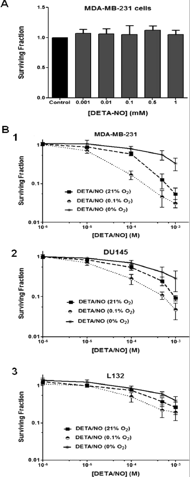
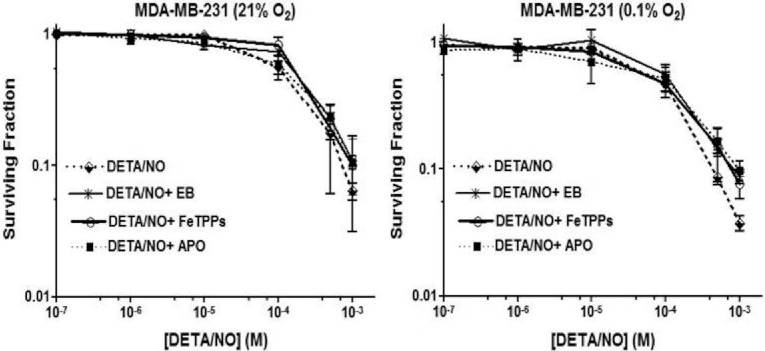
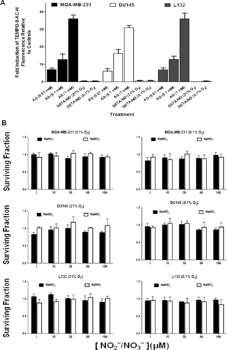
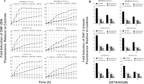
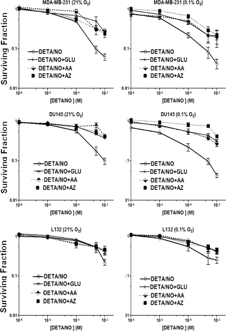
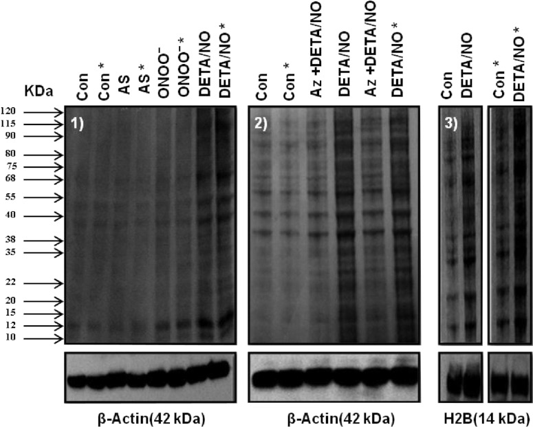
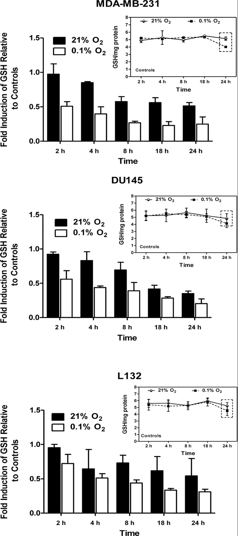
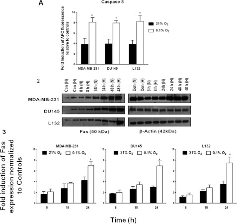
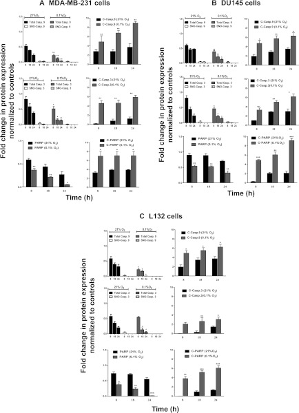
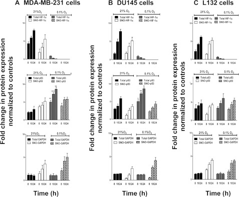
The relationship between the biological activity of NO and its chemistry is complex. The objectives of this study were to investigate the influence of oxygen tension on the cytotoxicity of the NO• donor DETA/NO and to determine the effects of oxygen tension on the key RNS (reactive nitrogen species) responsible for any subsequent toxicity. The findings presented in this study indicate that the DETA/NO-mediated cytotoxic effects were enhanced under hypoxic conditions. Further investigations revealed that neither ONOO⁻ (peroxynitrite) nor nitroxyl was generated. Fluorimetric analysis in the presence of scavengers suggest for the first time that another RNS, dinitrogen trioxide may be responsible for the cytotoxicity with DETA/NO. Results showed destabilization of HIF (hypoxia inducible factor)-1α and depletion of GSH levels following the treatment with DETA/NO under hypoxia, which renders cells more susceptible to DETA/NO cytotoxicity, and could account for another mechanism of DETA/NO cytotoxicity under hypoxia. In addition, there was significant accumulation of nuclear p53, which showed that p53 itself might be a target for S-nitrosylation following the treatment with DETA/NO. Both the intrinsic apoptotic pathway and the Fas extrinsic apoptotic pathway were also activated. Finally, GAPDH (glyceraldehyde-3-phosphate dehydrogenase) is another important S-nitrosylated protein that may possibly play a key role in DETA/NO-mediated apoptosis and cytotoxicity. Therefore this study elucidates further mechanisms of DETA/NO mediated cytotoxicity with respect to S-nitrosylation that is emerging as a key player in the signalling and detection of DETA/NO-modified proteins in the tumour microenvironment.


Figures

Figure 1
Figure 1. Evaluation of NO-induced toxicity from DETA/NO
(A) Clonogenic assay showing surviving fraction of MDA-MB-231 cells after 1 week incubation following treatment with a range of pre-incubated DETA/NO concentrations. DETA/NO was prepared in the DMEM medium and incubated at 37°C for 7 days to ensure complete liberation of NO molecules before addition to MDA-MB-231 cells for the duration of the assay (24 h). (B) Clonogenic assays to determine the effect of DETA/NO concentration on cell survival under normoxia, and 0.1% O2. MDA-MB-231, DU145 and L132 cells were incubated under either normoxic or hypoxic [21% (v/v) O2 or 0.1% (v/v) O2, 0% (v/v) O2] conditions for 24 h at 37°C prior to the 24 h treatment with DETA/NO. Fresh medium was placed on the cells, which were then incubated for 2 weeks and then stained with 0.4% crystal violet for colony counts. Data are the means of three independent experiments±S.E.M. Statistical significance was calculated using a one-way ANOVA. Exposure to DETA/NO under hypoxia at 0.1% (v/v) O2 was significantly more cytotoxic than in normoxia (P=0.0084, 0.0317, 0.0074, for MDA-MB-231, DU145 and L132 cells, respectively). Treatment of cells with DETA/NO under anoxia produced significantly less cytotoxicity compared with 0.1% hypoxia and normoxia (P=0.0045, 0.008, 0.046 under anoxia compared with normoxia for MDA-MB-231, DU145, and L132 cells, respectively; P=0.004, 0.0001, 0.001 under anoxia compared with 0.1% hypoxia for MDA-MB-231, DU145 and L132 cells, respectively, using one-way ANOVA test).
Figure 2
Figure 2. Clonogenic assay to determine the effects of the ONOO scavengers Ebselen (EB 10 μM), FeTPPs (50 μM) and Apocynin (APO 300 μM) on cell survival following the treatment with DETA/NO under normoxia and 0.1% (v/v) O2
MDA-MB-231 cells were incubated under either normoxic or hypoxic [21% (v/v) O2 or 0.1% (v/v) O2] conditions for 24 h at 37°C. ONOO scavengers were added to the cells 2 h prior to the 24 h treatment with DETA/NO. Fresh medium was placed on the cells, which were then incubated for 2 weeks and then stained with 0.4% crystal violet for colony counts. Data are the means of three independent experiments±S.E.M. Statistical significance was calculated using a one-way ANOVA.
Figure 3
Figure 3. Contribution of nitroxyl, nitrite and nitrate towards NO-mediated cytotoxicity
(A) TEMPO-9-AC was used to detect HNO following cellular exposure to DETA/NO. MDA-MB-231, DU145 and L132 cells were treated with LD50 concentrations of DETA/NO for 24 h under normoxic or 0.1% hypoxic conditions and the response compared with AS (10 μM–1 mM). Fluorescence was measured using a fluorescence plate reader with an excitation wavelength of 361 nm and a detection wavelength of 430 nm. Data were plotted as fold induction of TEMPO-9-AC-H relative to the controls. Data are the means of four independent experiments±S.E.M. (B) Clonogenic assays to determine the effect of NO2\NO3 on cellular survival under normoxia, and 0.1% (v/v) O2. Cells were incubated under either normoxic or 0.1% hypoxic conditions with different doses of NaNO2 and NaNO3 for 24 h at 37°C. Fresh medium was then placed on the cells, which were then incubated for 2 weeks and then stained with 0.4% crystal violet for colony counts. Data are the results of three independent experiments.
Figure 4
Figure 4. Measurement of N2O3 production from DETA/NO using a DAF-2DA probe
(A) Different DETA/NO concentrations (10 μM–1 mM) were exposed to MDA-MB-231, DU145 and L132 cells loaded with DAF-2DA probe under normoxia and 0.1% (v/v) O2. Fluorescence was measured using a fluorescence plate reader with an excitation wavelength of 485 nm and a detection wavelength of 530 nm. Data were plotted as fold induction of DAF-2 triazole relative to the controls. Data are the means of four independent experiments±S.E.M. (B) Quantification of N2O3 formation using DAF-2DA following the treatment of MDA-MB-231, DU145 and L132 cells with DETA/NO±N2O3 scavengers ascorbic acid (AA;1 mM), L-GSH reduced (GSH; 1 mM) and sodium azide (AZ;1 mM) under normoxia or 0.1% hypoxia. Data were plotted as fold induction of DAF-2 triazole fluorescence relative to the controls. Data are the means of three independent experiments±S.E.M. The asterisk indicates the significant difference in DAF-2 triazole fluorescence with DETA/NO±N2O3 scavengers compared with DETA/NO (*P≤0.05, **P≤0.005, ***P≤0.0005 using a two-tailed unpaired t test).
Figure 5
Figure 5. Dose–response curves showing survival of MDA-MB-231, DU145 and L132 cells following the exposure to various concentrations of DETA/NO, under both normoxic or 0.1% hypoxic conditions±N2O3 scavengers ascorbic acid (AA; 1 mM), L-GSH reduced (GSH; 1 mM) and sodium azide (AZ; 1 mM)
Data are the mean of three independent experiments±S.E.M. Statistical significance was calculated using a one-way ANOVA [P=0.003, 0.044, 0.0001 for MDA-MB-231, DU145 and L132 cells, respectively, treated with DETA/NO±sodium azide under 21% (v/v) O2 compared with DETA/NO treatments; P=0.0001 for MDA-MB-231, DU145 and L132 cells treated with DETA/NO±sodium azide under 0.1% (v/v) O2 compared with the DETA/NO treatments].
Figure 6
Figure 6. Biotin switch assay to determine the extent of protein S-nitrosylation following exposure to NO and ONOO
RSNO profile in MDA-MB-231 cells following the treatment with ONOO (1 mM), AS (1 mM), or LD50 concentrations of DETA/NO under normoxia or 0.1% hypoxia (*), (2) or MDA-MB-231 cells treated with DETA/NO±sodium azide. (3) The S-nitrosylated total protein profile in nuclear extracts following the treatment with LD50 concentrations of DETA/NO. Whole cell or nuclear lysates were subjected to the biotin switch method, biotin labelled nitrosoproteins were separated on SDS/PAGE (10% gel) and detected by Western blot analysis with an anti-biotin antibody. In parallel, β-actin or H2B protein levels served as a loading control. Images shown are the representative blots of three independent experiments. Arrows on the left point to visible bands representing the molecular mass of S-nitrosylated proteins.
Figure 7
Figure 7. NO suppression of glutathione
MDA-MB-231, DU145 and L132 cells were incubated under normoxia or 0.1% hypoxia for 2–24 h (line graphs) or treated with LD50 values of DETA/NO for 1–24 h at either normoxia or 0.1% hypoxia (bar graphs) and GSH content was determined using an optimized enzymatic recycling method that uses GR for GSH quantification. All data were normalized to mg protein, and DETA/NO bar graphs were expressed as fold induction of GSH/mg protein relative to controls. Data are the mean of four independent experiments±S.E.M.
Figure 8
Figure 8. Up-regulation of extrinsic apoptotic pathways following treatment with DETA/NO
(A) Caspase 8 activity was measured in MDA-MB-231, DU145 and L132 cells following treatment with LD50 values of DETA/NO for 24 h either at normoxia or 0.1% hypoxia using a fluorigenic substrate AFC as an indicator for caspase 8 activity. Data were plotted as fold induction of AFC fluorescence relative to controls. Data are the means of three independent experiments±S.E.M. (B) Detection of Fas receptor expression following the treatment of MDA-MB-231, DU145 and L132 cells with LD50 concentrations of DETA/NO. Data were plotted as fold change in Fas expression normalized to controls over a time course for both 21% O2 normoxia (N) and 0.1% O2 hypoxic conditions (H). Representative Western blots and the mean densitometric values±S.E.M. for three independent experiments are shown.
Figure 9
Figure 9. Evidence of activation of intrinsic apoptotic proteins following treatment with DETA/NO
Caspase 9 and 3 (total and S-nitrosylated) and PARP cleavage over a time course following the treatment of MDA-MB-231 (A), DU145 (B) and L132 (C) cells with LD50 concentrations of DETA/NO under both normoxia and 0.1% (v/v) O2 hypoxia. Data are the mean densitometric values±S.E.M. for three independent experiments and are plotted as fold change in protein expression normalized to controls over a time course of both 21% O2 normoxia and 0.1% (v/v) O2 hypoxia. The asterisk indicates significant difference under 0.1% (v/v) O2 hypoxia compared with normoxia for the total protein (*P≤0.05, **P≤0.005, ***P≤0.0005 using a two-tailed an unpaired t test).
Figure 10
Figure 10. Western blot analysis of nuclear proteins (HIF-1α, p53 and GAPDH) expression over time under both normoxia and 0.1% O2 hypoxia
(A) MDA-MB-231, (B) DU145 and (C) L132 cells. Both S-nitrosylated and total proteins were examined as previously mentioned in Figure 9. Data were the mean densitometric values±S.E.M. for three independent experiments and was plotted as fold change in protein expression normalized to controls over a time course of both 21% (v/v) O2 normoxia and 0.1% (v/v) O2 hypoxia.

Similar articles

Cited by

References

    1. Lancaster J. A tutorial on the diffusibility and reactivity of free nitric oxide. Nitric Oxide. 1997;1:18–30. - PubMed
    1. Muntané J., De la Mata M. Nitric oxide and cancer. World J. Hepatol. 2010;2:337–345. - PMC - PubMed
    1. Ying L., Hofseth L. J. An emerging role for endothelial nitric oxide synthase in chronic inflammation and cancer. Cancer Res. 2007;67:1407–1415. - PubMed
    1. Hirst D., Robson T. Targeting nitric oxide for cancer therapy. J. Pharm. Pharmacol. 2007;59:3–13. - PubMed
    1. Shami P. J., Saavedra J. E., Bonifant C. L., Chu J., Udupi V., Malaviya S., Carr B. I., Kar S., Wang M., Jia L. Antitumor activity of JS-K [O2-(2, 4-dinitrophenyl) 1-[(4-ethoxycarbonyl) piperazin-1-yl] diazen-1-ium-1, 2-diolate] and related O2-aryl diazeniumdiolates in vitro and in vivo. J. Med. Chem. 2006;49:4356–4366. - PubMed

MeSH terms