. 2013 Mar 26;7(3):1961-73.
doi: 10.1021/nn304439f. Epub 2013 Feb 22.

Nanoparticle geometry and surface orientation influence mode of cellular uptake

Affiliations

Nanoparticle geometry and surface orientation influence mode of cellular uptake

Heather Herd et al. ACS Nano. .

Abstract

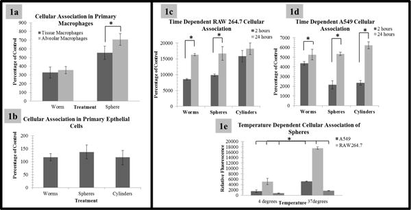
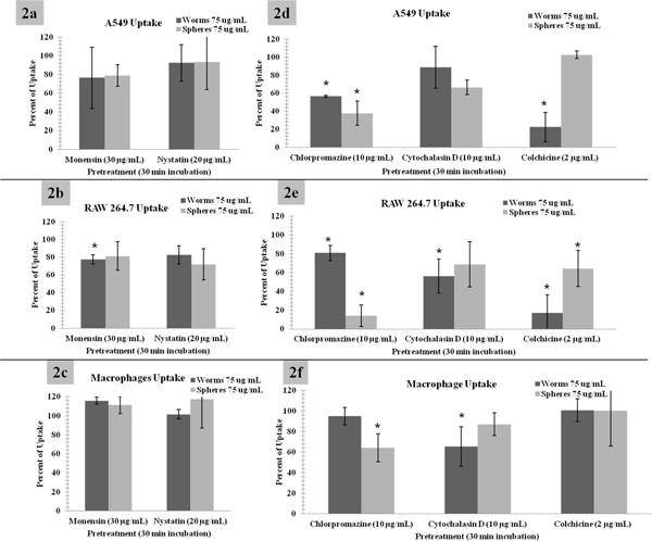
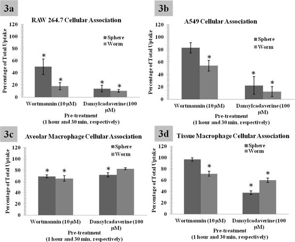
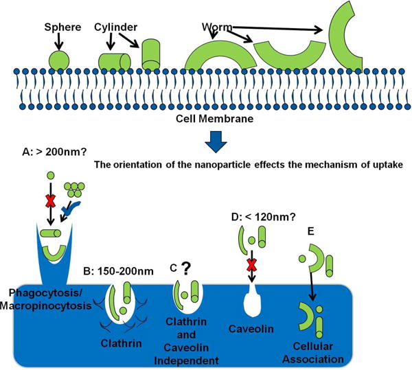
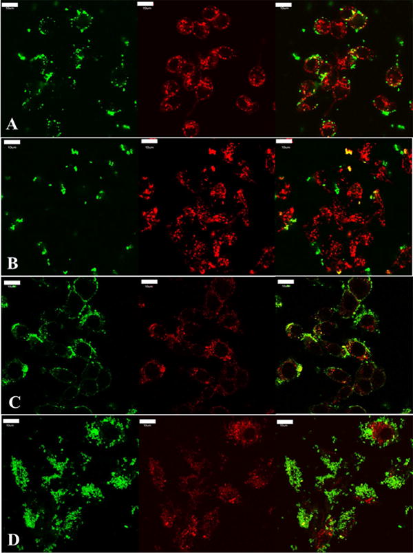
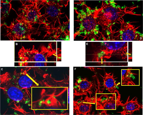
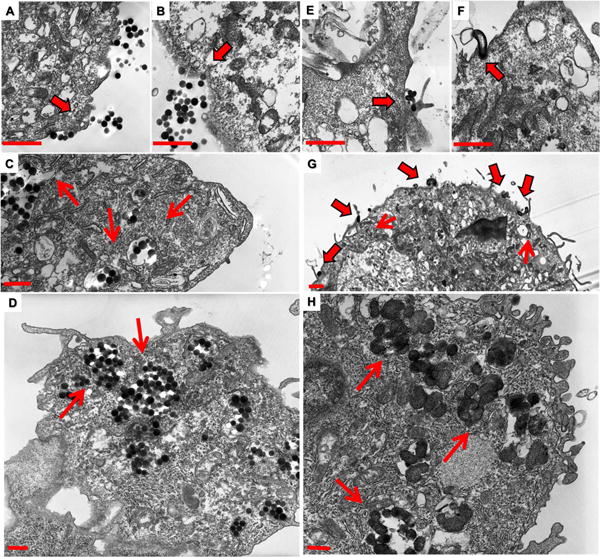
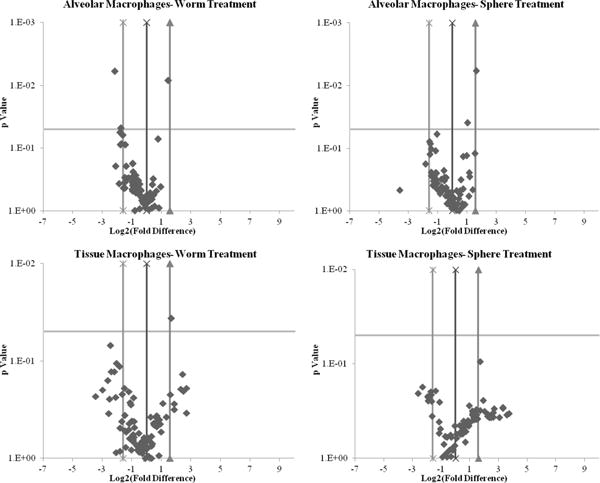
In order to engineer safer nanomaterials, there is a need to understand, systematically evaluate, and develop constructs with appropriate cellular uptake and intracellular fates. The overall goal of this project is to determine the uptake patterns of silica nanoparticle geometries in model cells, in order to aid in the identification of the role of geometry on cellular uptake and transport. In our experiments we observed a significant difference in the viability of two phenotypes of primary macrophages; immortalized macrophages exhibited similar patterns. However, both primary and immortalized epithelial cells did not exhibit toxicity profiles. Interestingly uptake of these geometries in all cell lines exhibited very different time-dependent patterns. A screening of a series of chemical inhibitors of endocytosis was performed to isolate the uptake mechanisms of the different particles. The results show that all geometries exhibit very different uptake profiles and that this may be due to the orientation of the nanoparticles when they interact with the cell surface. Additionally, evidence suggests that these uptake patterns initialize different downstream cellular pathways, dependent on cell type and phenotype.


Figures

Figure 1
Figure 1. Basal uptake of geometrically defined nanoparticles
Relative uptake of 75 μg/mL of nanoparticles at specified time points assessed via FACS. In 1a, macrophages exhibit significant uptake. However, both the phenotype of the macrophage and the geometry (at this time point, 1.5 hours) appear to play important roles in the degree of uptake. It is also important to note that alveolar macrophages treated with spherical nanoparticles when compared to worm like nanoparticles appear to have a greater degree of nanoparticle uptake, at this time point). This suggests a phenotypic and geometric implication. 1b. Epithelial cells exhibit little to no nanoparticle uptake when compared to control. 1c and 1d: Relative uptake of 75 μg/mL of nanoparticles at specified time points assessed via FACS. As shown, initial time point analysis shows a variation in the uptake rate of nanoparticles, dependent upon geometry while later time points appear to have an equivalent rate of cellular association (for clarity some time points have been removed, please see Supplemental Figure 5 for all time points). 1e: Representative graph of the relative uptake of spherical nanoparticles as a function of temperature in model cells. Alveolar macrophages are depicted by their increased uptake potential. The graph provides confirmation of energy dependent mechanisms of uptake. Please note: graphs are represented as percentage of control or the background provided by FACS analysis of cells incubated without nanoparticles; so 100% would be 100% of control. Low levels of autofluorescence were indicated for immortalized lines, while high levels were indicated for primary cells due to donor variations including unknown patient treatment (i.e. chemotherapeutics, smoker or non-smoker, other diseases/treatments, etc.) *Indicates statistical significance p value < 0.05
Figure 2
Figure 2
2a, 2b, and 2c: Relative level of nanoparticle uptake when incubated with Monensin (clathrin and caveolin independent endocytosis inhibitor) and Nystatin (caveolin endocytosis inhibitor). This suggests that caveolin mediated endocytosis is not involved in the internalization of these nanoparticle systems. 2d, 2e, and 2f: Cytochalasin D and colchicine are phagocytic and macropinocytosis inhibitors, while chlorpromazine inhibits clathrin mediated mechanisms. The results show that there is a statistically significant reduction in nanoparticle uptake for these three inhibitors, but all geometries show very different uptake profiles. This suggests that the mechanisms by which these particles are entering the cells vary and are dependent on the relative shape (or size). For clarity and due to the significant similarities of worms and cylinders, cylindrical data has been moved to the supplemental materials. Please note: graphs are represented as percentage of uptake or the background provided by FACS analysis of cells incubated with spheres or worms without the respective inhibitor. *Indicates statistical significance from control p value < 0.05. Macrophages are alveolar macrophages.
Figure 3
Figure 3
Flow cytometry analysis to quantitatively assess the uptake of nanoparticles. Due to low specificity in endocytic inhibitors, dansylcadaverine (a clathrin inhibitor) and wortmannin (a phagocytic and macropinocyotsis inhibitor) were utilized to confirm the apparent variations in uptake due to changes in geometry. As shown, dansylcadaverine blocks both sphere and worm uptake, but to a more significant degree spherical uptake. However, wortmannin is observed to block worm like uptake to a more significant degree, supporting geometric variations in cellular uptake. Additionally, differences in uptake due to the cell phenotype were observed. For clarity and due to the significant similarities of worms and cylinders, cylindrical data has been moved to the supplemental materials. Please note: graphs are represented as percentage of uptake or the background provided by FACS analysis of cells incubated with spheres or worms without the respective inhibitor. *Indicates statistical significance from control p value < 0.05.
Figure 4
Figure 4
A cartoon depicting the hypothesis that the orientation of the nanoparticle influences the mechanism of uptake. A: Macropinocytosis has been shown to occur at the micro scale, fitting with one dimension of cylinders and worms. However, spheres, unless aggregated do not fit this criteria. B: Theoretically if oriented properly all particles could be uptaken via clathrin mediated mechanisms. C and E: Other mechanisms of uptake not readily identifiable are plausible. D: Caveolin mediated invaginations are theoretically much too small for the nanoparticle size range tested here.
Figure 5
Figure 5
Transferrin and dextran, intracellular markers of clathrin mediated endocytosis and fluid phase endocytosis, respectively, were co-incubated with silica nanoparticles to investigate the degree of co-localization to confirm clathrin and fluid phase mediated mechanisms; RAW 264.7 cells shown. A and B) Depicted is a single focal plane and the respective channels of live cells after 15 mins of incubation. Transferrin is labeled in red, nanoparticles are labeled in green (A is spherical treatment and B is worm like treatment both at 75 μg/mL), and the co-localization is depicted in yellow between particles and transferrin. C and D) Depicted is the fluorescence from all Z stacks and respective channels of fixed cells after 30 mins of incubation. Dextran is labeled in red, particles are labeled in green (C is spherical treatment and D is worm like treatment at 75 μg/mL), and yellow represents the co-localization between nanoparticles and dextran. There appears to be a higher degree of co-localization with both these cellular internalization markers with spherical nanoparticles when compared to worm like nanoparticles, suggesting that a higher degree of clathrin and fluid phase mediated endocytosis occurs with these systems. It is important to note that the co-localization (yellow) in all images, suggests that clathrin and fluid phase mediated endocytosis is at play for all geometries tested. For clarity and due to the significant similarities of worms and cylinders, cylindrical data has been moved to the supplemental materials. Scale bar 10 μm.
Figure 6
Figure 6
Actin polymerization staining in RAW 264.7 cells, involved in mechanisms of endocytosis is depicted to visualize the hallmark endocytic process involved in the internalization of these particles. Red: phalloidin stain of actin polymerization, green: nanoparticle FITC attachment, and blue: DAPI nucleus stain. A, B and C) Spherical nanoparticle and D, E and F) worm nanoparticle treatment after 15 mins. Spherical nanoparticle treatment (A) and worm nanoparticle treatment (D) appears to induce very different polymerization patterns. The polymerization patterns observed in treated cells include invaginations within the membrane associated with nanomaterials, identified in the zoomed insert in C and in the depiction of the z stack in B. While other polymerization patterns appear to be extravasations from the membrane associated with nanomaterials, marked in the zoomed inserts in F and the z stack in E. These polymerization patterns could suggest the involvement of clathrin mediated, macropinocytic and phagocytic mechanisms.). For clarity and due to the significant similarities of worms and cylinders, cylindrical data has been moved to the supplemental materials.
Figure 7
Figure 7
Following 15 mins of incubation with both worm and spherical nanoparticles, cells were fixed and imaged via TEM. Membrane invaginations are associated with spherical particles (A and B). Membrane extravasations however are observed to be associated with worm particles (E and G) and membrane wrapping associated with worm like particles (F and G) are observed. Both nanoparticles are observed within the cytoplasm of these cells at 15 mins (C and G) while there is a greater degree of uptake of spherical nanoparticles at this time point. This suggests that spherical particles are taken up more rapidly than worm nanoparticles. However, a greater amount of both nanoparticles are internalized at 24 hour time points, as we observed previously (D and H). Additionally, there appears to be some type of sequestering mechanism at play.). For clarity and due to the significant similarities of worms and cylinders, cylindrical data has been moved to the supplemental materials. Scale bars 1μm.
Figure 8
Figure 8
PI3-kinase array analysis volcano plots of primary macrophages regulated genes. More regulation is observed in tissue macrophages when compared to alveolar macrophages suggesting a different pattern of uptake due to phenotypic differences. This could also potentially play an important role in toxicity. Additionally, worms regulate genes to a more significant extent than spheres do in both tissue and alveolar macrophages. However, it is plausible that particle treatment itself could induce this regulation.

Similar articles

Cited by

References

    1. Nel AE, Madler L, Velegol D, Xia T, Hoek EM, Somasundaran P, Klaessig F, Castranova V, Thompson M. Understanding Biophysicochemical Interactions at the Nano-Bio Interface. Nat Mater. 2009;8:543–557. - PubMed
    1. Jones AT, Gumbleton M, Duncan R. Understanding Endocytic Pathways and Intracellular Trafficking: A Prerequisite for Effective Design of Advanced Drug Delivery Systems. Adv Drug Delivery Rev. 2003;55:1353–1357. - PubMed
    1. Wang LH, Rothberg KG, Anderson RG. Mis-Assembly of Clathrin Lattices on Endosomes Reveals a Regulatory Switch for Coated Pit Formation. J Cell Biol. 1993;123:1107–1117. - PMC - PubMed
    1. Pelkmans L, Burli T, Zerial M, Helenius A. Caveolin-Stabilized Membrane Domains as Multifunctional Transport and Sorting Devices in Endocytic Membrane Traffic. Cell. 2004;118:767–780. - PubMed
    1. Wiewrodt R, Thomas AP, Cipelletti L, Christofidou-Solomidou M, Weitz DA, Feinstein SI, Schaffer D, Albelda SM, Koval M, Muzykantov VR. Size-Dependent Intracellular Immunotargeting of Therapeutic Cargoes into Endothelial Cells. Blood. 2002;99:912–922. - PubMed

Publication types

MeSH terms