. 2013 Feb 12:13:11.
doi: 10.1186/1472-6750-13-11.

Stable nuclear transformation of Eudorina elegans

Affiliations

Stable nuclear transformation of Eudorina elegans

Kai Lerche et al. BMC Biotechnol. .

Abstract

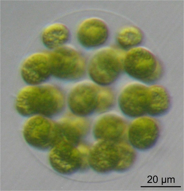
Background: A fundamental step in evolution was the transition from unicellular to differentiated, multicellular organisms. Volvocine algae have been used for several decades as a model lineage to investigate the evolutionary aspects of multicellularity and cellular differentiation. There are two well-studied volvocine species, a unicellular alga (Chlamydomonas reinhardtii) and a multicellular alga with differentiated cell types (Volvox carteri). Species with intermediate characteristics also exist, which blur the boundaries between unicellularity and differentiated multicellularity. These species include the globular alga Eudorina elegans, which is composed of 16-32 cells. However, detailed molecular analyses of E. elegans require genetic manipulation. Unfortunately, genetic engineering has not yet been established for Eudorina, and only limited DNA and/or protein sequence information is available.

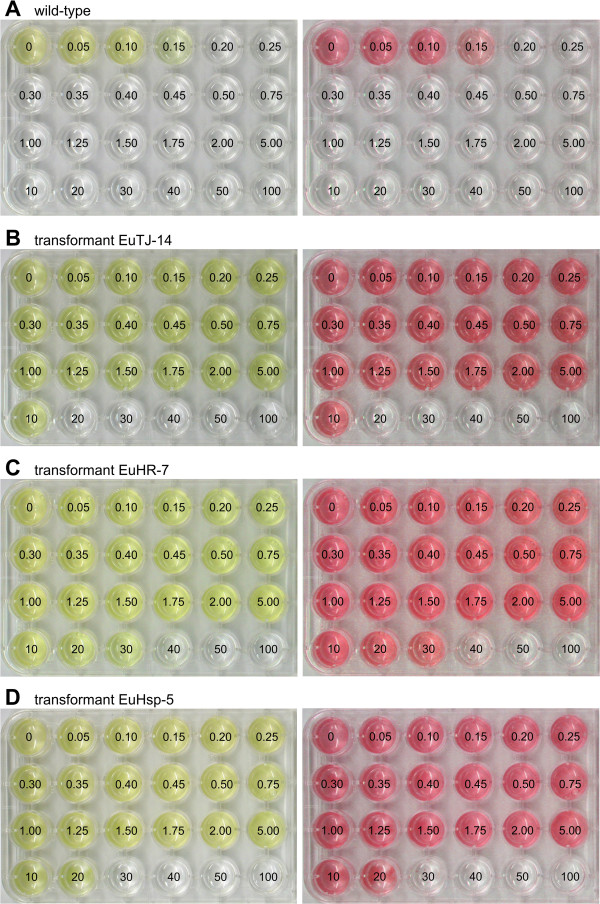
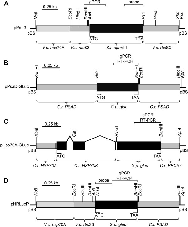
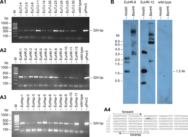
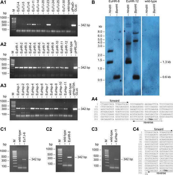
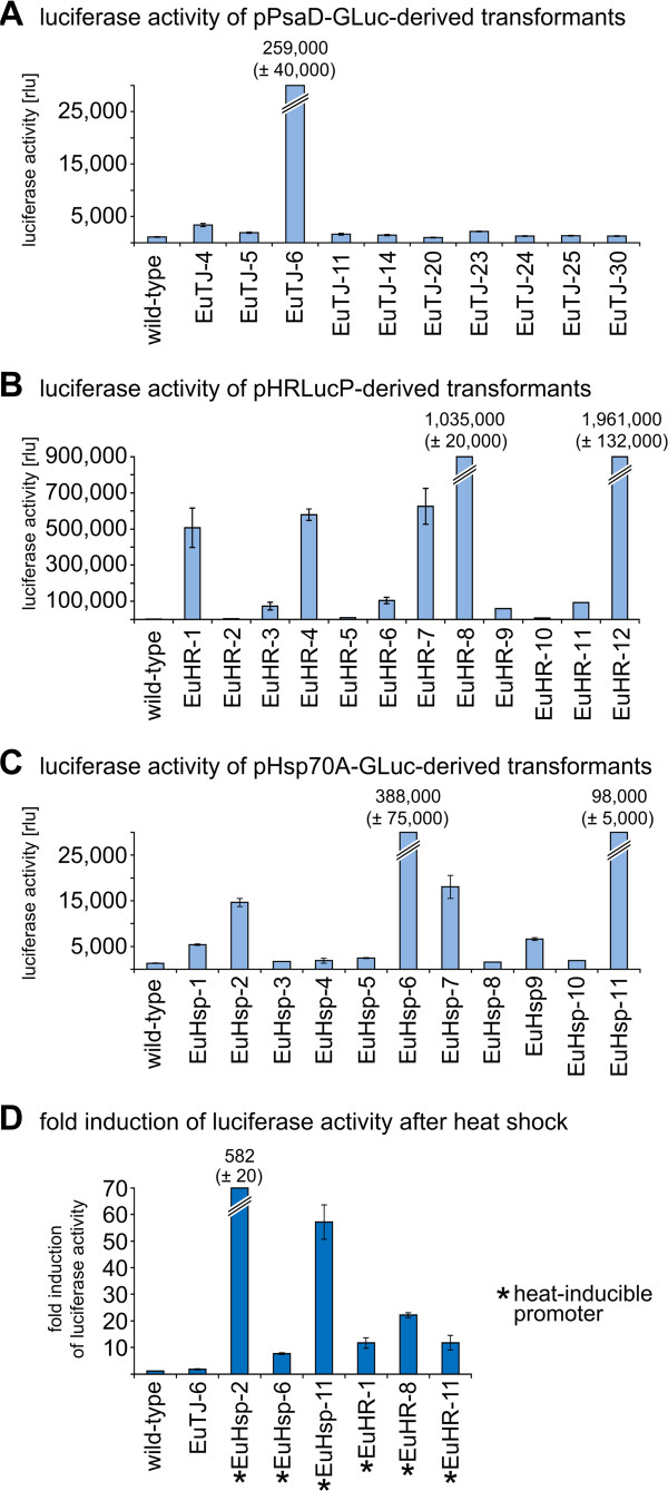
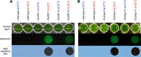
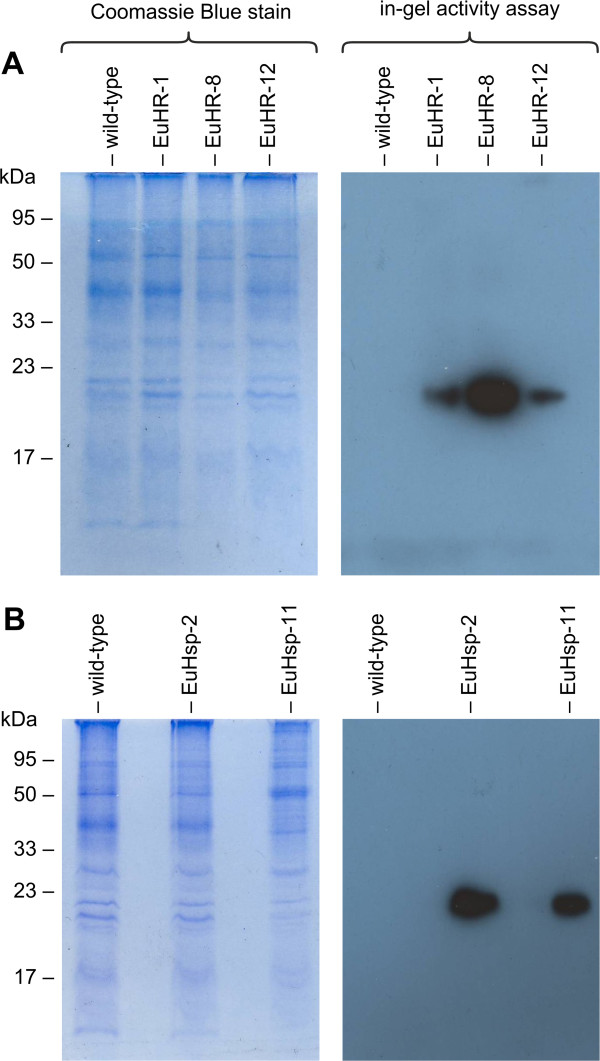
Results: Here, we describe the stable nuclear transformation of E. elegans by particle bombardment using both a chimeric selectable marker and reporter genes from different heterologous sources. Transgenic algae resistant to paromomycin were achieved using the aminoglycoside 3'-phosphotransferase VIII (aphVIII) gene of Streptomyces rimosus, an actinobacterium, under the control of an artificial promoter consisting of two V. carteri promoters in tandem. Transformants exhibited an increase in resistance to paromomycin by up to 333-fold. Co-transformation with non-selectable plasmids was achieved with a rate of 50 - 100%. The luciferase (gluc) gene from the marine copepod Gaussia princeps, which previously was engineered to match the codon usage of C. reinhardtii, was used as a reporter gene. The expression of gluc was mediated by promoters from C. reinhardtii and V. carteri. Heterologous heat shock promoters induced an increase in luciferase activity (up to 600-fold) at elevated temperatures. Long-term stability and both constitutive and inducible expression of the co-bombarded gluc gene was demonstrated by transcription analysis and bioluminescence assays.

Conclusions: Heterologous flanking sequences, including promoters, work in E. elegans and permit both constitutive and inducible expression of heterologous genes. Stable nuclear transformation of E. elegans is now routine. Thus, we show that genetic engineering of a species is possible even without the resources of endogenous genes and promoters.


Figures

Figure 1
Figure 1
The wild-type phenotype of E. elegans. A vegetatively grown spheroid of E. elegans with ~32 biflagellate cells at the surface of the organism is shown. Each round-shaped cell contains a single large, cup-shaped chloroplast. Most of the spheroid volume consists of a complex but transparent glycoprotein-rich extracellular matrix that holds all of the cells in place.
Figure 2
Figure 2
Analysis of paromomycin resistance in wild-type and transgenic E. elegans strains. For analysis of antibiotic resistance, identical quantities of wild-type or transgenic Eudorina cells were exposed to increasing concentrations of paromomycin, and incubated for 10 days. Numbers refer to the concentration of paromomycin [μg/ml] utilized. Natural color (left) and red-shifted, false-color images (right) are shown. (A) Wild-type E. elegans strain UTEX 1193 used as a reference control. (B-D) Transgenic E. elegans strains co-transformed with pPmr3, the selectable marker plasmid, in addition to a second, non-selectable reporter gene plasmid. (B) Transformant EuTJ-14 was co-transformed with the plasmids pPmr3 and pPsaD-GLuc. (C) Transformant EuHR-7 was co-transformed with the plasmids pPmr3 and pHRLucP. (D) Transformant EuHsp-5 was co-transformed with the plasmids pPmr3 and pHsp70A-GLuc.
Figure 3
Figure 3
Schematic diagram of the selectable marker plasmid and co-transformed,non-selectable plasmids. (A) The chimeric selectable marker plasmid pPmr3. (B-D) Co-transformed, non-selectable plasmids containing reporter genes: (B) pPsaD-GLuc, (C) pHsp70A-GLuc, and (D) pHRLucP. The angled lines in (C) indicate introns. (A-D) Amplified genomic (gPCR) or RT-PCR fragments as well as probes used for Southern blots are indicated. V.c., Volvox carteri; C.r., Chlamydomonas reinhardtii; S.r., Streptomyces rimosus; G.p., Gaussia princeps; gluc, luciferase gene, pBS, pBluescript II vector.
Figure 4
Figure 4
Detection of the aph VIII gene in transformants. (A1-A3) Paromomycin-resistant transformants and the parental wild-type strain were analyzed for presence of the aphVIII gene in the genome by genomic PCR. The expected size of the aphVIII fragment produced in transformants was 324 bp (Figure 3A). The rightmost lane (pPmr3) shows a positive control using pPmr3 plasmid DNA as the template. Lane M refers to the molecular weight marker. (A4) Sequence obtained from the amplified and cloned aphVIII fragments. The positions of the primers and the start codon (bold) are indicated. (B) Southern blot analysis of HindIII- or BamHI-digested genomic DNA of two randomly chosen transformants, EuHR-8 and EuHR-12, and from the parental wild-type strain. The blot was probed using a 282-bp fragment of the aphVIII coding region (Figure 3A).
Figure 5
Figure 5
Demonstration of co-transformation and transcription of heterologous genes. (A1-A3) Paromomycin-resistant transformants were analyzed for the presence of the co-transformed, non-selectable DNA in the genome. PCRs were conducted using genomic DNA from transformants co-transformed with the pPsaD-GLuc (A1), pHRLucP (A2), or pHsp70A-GLuc (A3) plasmids as template. The parental wild-type strain was analyzed as a control. Primers were specific for the heterologous gluc gene, and a 342-bp PCR fragment (Figure 3B–D) was expected in co-transformants. The rightmost lane shows a positive control using DNA of the co-transformed plasmid as the template. Lane M refers to the molecular weight marker. (A4) Sequence obtained from the amplified and cloned gluc fragments. Primer positions, the stop codon (bold), and a BamHI restriction site (italics) are indicated. (B) Southern blot analysis of HindIII- or BamHI-digested genomic DNA from transformants EuHR-8 and EuHR-12, which were produced using the pPmr3 and pHRLucP plasmids, and from the parental wild-type strain. The blot was probed using a 258-bp fragment of the gluc gene (Figure 3D). (C1-C3) Transcription analysis by RT-PCR. RNA from paromomycin-resistant transformants co-bombarded with the non-selectable plasmids pPsaD-GLuc (C1), pHRLucP (C2), or pHsp70A-GLuc (C3) was reverse transcribed, and products were amplified by PCR using gluc-specific primers. Co-transformants were expected to yield a 342-bp cDNA fragment of the gluc gene (Figure 3B–D). The parental wild-type strain was used as a control. (C4) Sequence obtained from the cloned gluc cDNA fragments. Primer positions, the stop codon (bold), and a BamHI restriction site (italics) are indicated. Lane M refers to the molecular weight marker.
Figure 6
Figure 6
Quantification and inducibility of luciferase activity in transformants expressing the luciferase gene. Luciferase activity in transformants was assayed using a luminometer. The bars represent the mean of three independent experiments. The standard deviation is indicated. (A) Luciferase activity of pPsaD-GLuc-derived transformants (EuTJ…) compared to the wild-type strain. (B) Luciferase activity of pHRLucP-derived transformants (EuHR…) compared to the wild-type strain. (C) Luciferase activity of pHsp70A-GLuc-derived transformants (EuHsp…) compared to the wild-type strain. (D) Fold induction of luciferase activity in heat-shocked transformants (42°C) compared to non-heat-shocked transformants. In pPsaD-GLuc-derived transformants (EuTJ…), the luciferase gene is driven by the Chlamydomonas reinhardtii psaD promoter, and in pHRLucP-derived transformants (EuHR…), the luciferase is driven by the Volvox carteri hsp70A/rbcS3 tandem promoter. For pHsp70A-GLuc-derived transformants (EuHsp…), the luciferase gene is driven by the Chlamydomonas reinhardtii HSP70A promoter. Transformants with a gluc gene driven by a heat-inducible promoter are indicated (*). The parental wild-type strain was analyzed as a control. Transformants and wild-type colonies were subjected to a temperature shift from 27°C to 42°C for 1 h. After a 15 min recovery phase at 27°C, cells were lysed, and luciferase activity was assayed. As a reference control, non-heat-shocked transformants were analyzed in an identical fashion.
Figure 7
Figure 7
Visualization of inducible luciferase activity. Luciferase assay results for parental wild-type colonies and for several transformants with or without heat shock. (A) Transformants EuHR-1 and EuHR-12, generated with pHRLucP as the co-bombarded plasmid. (B) Transformants EuHsp-2 and EuHsp-11, generated with pHsp70A-GLuc as the co-bombarded plasmid. Algae cultures were divided into two aliquots; one aliquot was subjected to a heat shock at 42°C for 1 h, and the other aliquot was maintained at 27°C. Upper row: standard photo showing the assay setup. Middle row: photo without extraneous light in the darkroom following the addition of the coelenterazine substrate. Lower row: exposure to a light-sensitive film.
Figure 8
Figure 8
Detection of luciferase activity by in-gel activity assays. Cell extracts of heat-shocked transformants were subjected to SDS-PAGE. Subsequently, in-gel renaturation was performed, the coelenterazine-substrate was added, and the gel was exposed to a light-sensitive film (right panels). As a loading control, the same extracts were also stained with Coomassie Blue following SDS-PAGE (left panels). (A) Analysis of transformants EuHR-1, EuHR-8, and EuHR-12, which were generated with pHRLucP as the co-bombarded plasmid. (B) Analysis of transformants EuHsp-2 and EuHsp-11, generated with pHsp70A-GLuc as the co-bombarded plasmid. In each experiment, the parental wild-type strain was analyzed as a control.

Similar articles

Cited by

References

    1. Baldauf SL. The deep roots of eukaryotes. Science. 2003;300(5626):1703–1706. doi: 10.1126/science.1085544. - DOI - PubMed
    1. King N. The unicellular ancestry of animal development. Dev Cell. 2004;7(3):313–325. doi: 10.1016/j.devcel.2004.08.010. - DOI - PubMed
    1. Grosberg RK, Strathmann R. The evolution of multicellularity: a minor major transition? Annu Rev Ecol Evol Syst. 2007;38:621–654. doi: 10.1146/annurev.ecolsys.36.102403.114735. - DOI
    1. Prochnik SE, Umen J, Nedelcu AM, Hallmann A, Miller SM, Nishii I, Ferris P, Kuo A, Mitros T, Fritz-Laylin LK. et al.Genomic analysis of organismal complexity in the multicellular green alga Volvox carteri. Science. 2010;329(5988):223–226. doi: 10.1126/science.1188800. - DOI - PMC - PubMed
    1. Kirk DL. Volvox: molecular-genetic origins of multicellularity and cellular differentiation. 1. Cambridge University Press, Cambridge; 1998.

Publication types

LinkOut - more resources