

Neuroprotection of rat retinal ganglion cells mediated through alpha7 nicotinic acetylcholine receptors
- PMID: 23402849
- PMCID: PMC3609885
- DOI: 10.1016/j.neuroscience.2013.02.003
Neuroprotection of rat retinal ganglion cells mediated through alpha7 nicotinic acetylcholine receptors
Abstract
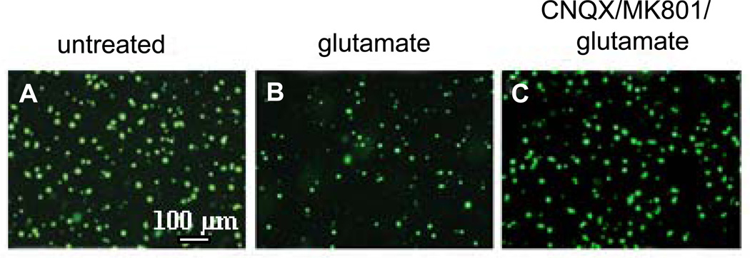
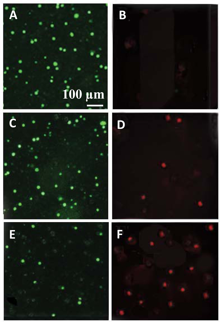
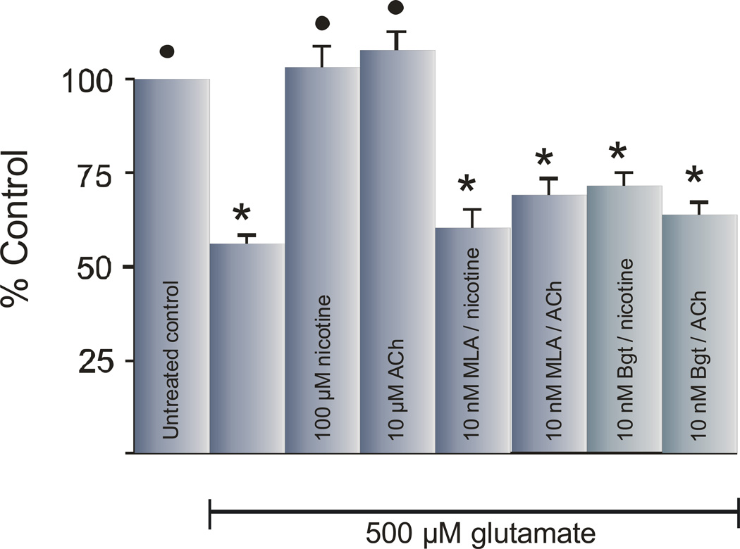
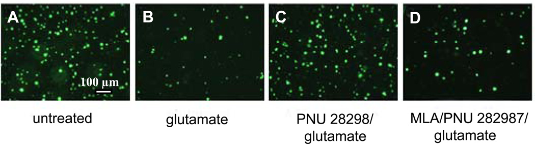
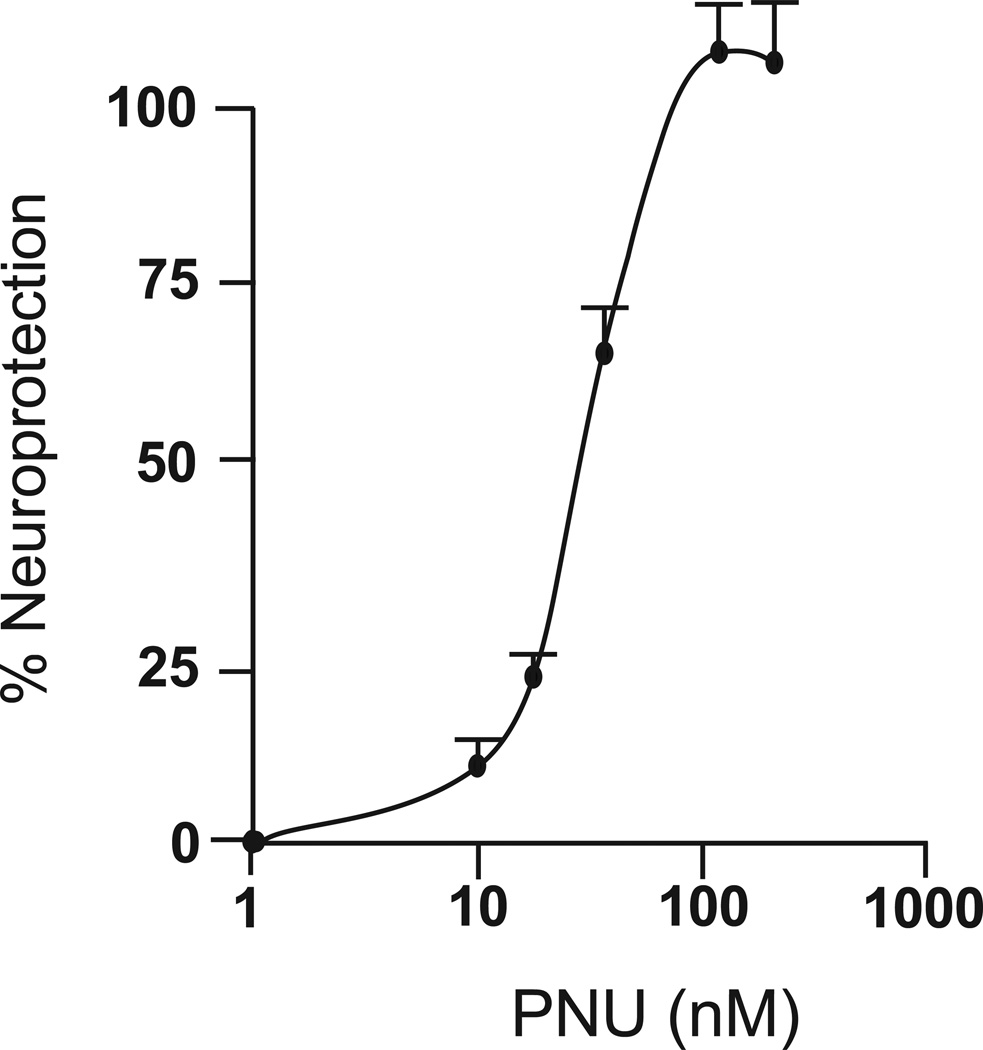
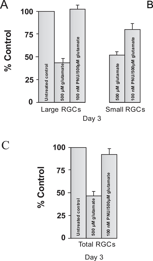
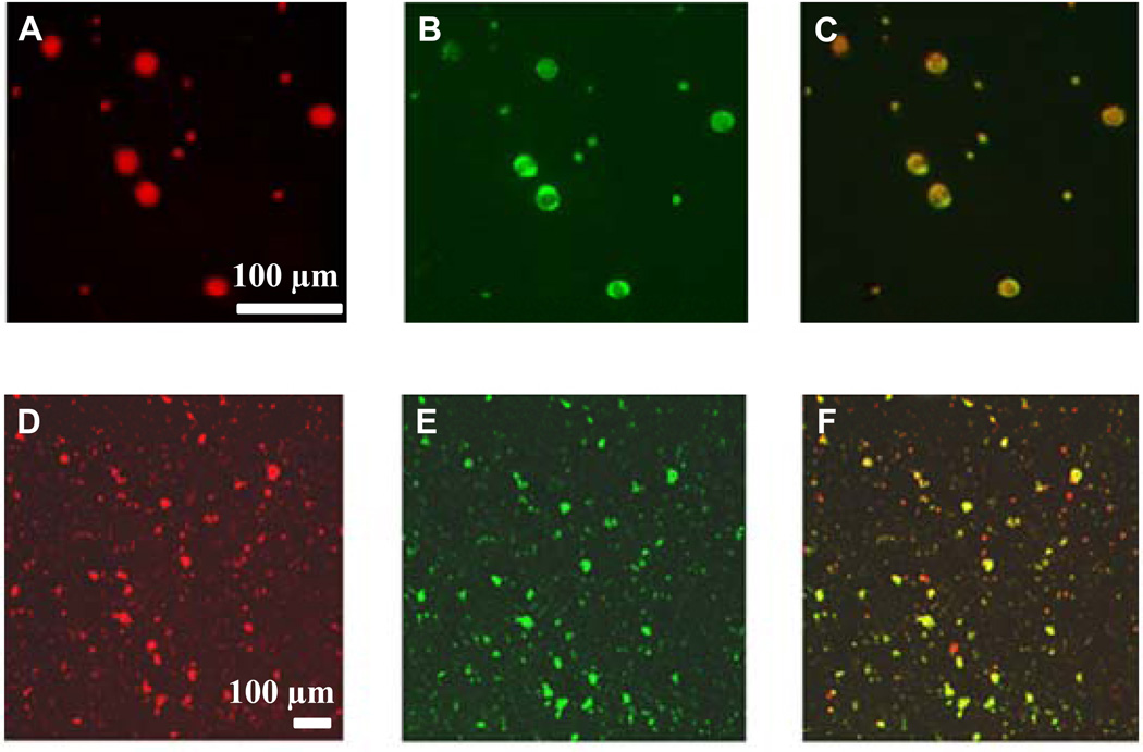
Glutamate-induced excitotoxicity is thought to play an important role in several neurodegenerative diseases in the central nervous system (CNS). In this study, neuroprotection against glutamate-induced excitotoxicity was analyzed using acetylcholine (ACh), nicotine and the α7 specific nicotinic acetylcholine receptor (α7 nAChR) agonist, N-[(3R)-1-azabicyclo[2.2.2]oct-3-yl]-4-chlorobenzamide hydrochloride (PNU-282987), in cultured adult rat retinal neurons. Adult Long Evans rat retinas were dissociated and retinal ganglion cells (RGCs) were isolated from all other retinal tissue using a two-step panning technique. Once isolated, RGCs were cultured under various pharmacological conditions to demonstrate excitotoxicity and neuroprotection against excitotoxicity. After 3 days, RGCs were immunostained with antibodies against the glycoprotein, Thy 1.1, counted and cell survival was assessed relative to control untreated conditions. 500 μM glutamate induced excitotoxicity in large and small RGCs in an adult rat dissociated culture. After 3 days in culture with glutamate, the cell survival of large RGCs decreased by an average of 48.16% while the cell survival of small RGCs decreased by an average of 42.03%. Using specific glutamate receptor agonists and antagonists, we provide evidence that the excitotoxic response was mediated through α-amino-3-hydroxy-5-methyl-4-isoxazolepropionic acid (AMPA)/kainic acid (KA) and N-methyl-d-aspartate (NMDA) glutamate receptors through an apoptotic mechanism. However, the excitotoxic effect of glutamate on all RGCs was eliminated if cells were cultured for an hour with 10 μM ACh, 100 μM nicotine or 100 nM of the α7 nAChR agonist, PNU-282987, before the glutamate insult. Inhibition studies using 10nM methyllycaconitine (MLA) or α-bungarotoxin (α-Bgt) supported the hypothesis that neuroprotection against glutamate-induced excitotoxicity on rat RGCs was mediated through α7 nAChRs. In immunocytochemical studies, double-labeled experiments using antibodies against Thy 1.1 and α7 nAChR subunits demonstrated that both large and small RGCs contained α7 nAChR subunits. The data presented in this study support the hypothesis that ACh and nicotinic acetylcholine receptor (nAChR) agonists provide neuroprotection against glutamate-induced excitotoxicity in adult rat RGCs through activation of α7 nAChR subunits. These studies lay the groundwork required for analyzing the effect of specific α7 nAChR agonists using in vivo models of excitotoxicity. Understanding the type of ACh receptors involved in neuroprotection in the rat retina could ultimately lead to therapeutic treatment for any CNS disease that involves excitotoxicity.
Copyright © 2013 IBRO. Published by Elsevier Ltd. All rights reserved.
Conflict of interest statement
The authors state no conflict of interest.
Figures
Similar articles
-
Tropisetron as a neuroprotective agent against glutamate-induced excitotoxicity and mechanisms of action.Neuropharmacology. 2013 Oct;73:111-21. doi: 10.1016/j.neuropharm.2013.05.020. Epub 2013 May 29. Neuropharmacology. 2013. PMID: 23727438 Free PMC article.
-
Acetylcholine neuroprotection against glutamate-induced excitotoxicity in adult pig retinal ganglion cells is partially mediated through alpha4 nAChRs.Exp Eye Res. 2006 Nov;83(5):1135-45. doi: 10.1016/j.exer.2006.05.022. Epub 2006 Aug 22. Exp Eye Res. 2006. PMID: 16928373
-
Acetylcholine protection of adult pig retinal ganglion cells from glutamate-induced excitotoxicity.Invest Ophthalmol Vis Sci. 2004 May;45(5):1531-43. doi: 10.1167/iovs.03-0406. Invest Ophthalmol Vis Sci. 2004. PMID: 15111612
-
Mechanisms of neuroprotective effects of nicotine and acetylcholinesterase inhibitors: role of alpha4 and alpha7 receptors in neuroprotection.J Mol Neurosci. 2010 Jan;40(1-2):211-6. doi: 10.1007/s12031-009-9236-1. J Mol Neurosci. 2010. PMID: 19714494 Review.
-
Advances in the discovery of novel positive allosteric modulators of the alpha7 nicotinic acetylcholine receptor.Recent Pat CNS Drug Discov. 2007 Jun;2(2):99-106. doi: 10.2174/157488907780832751. Recent Pat CNS Drug Discov. 2007. PMID: 18221220 Review.
Cited by
-
Alpha7 nicotinic acetylcholine receptor agonist promotes retinal ganglion cell function via modulating GABAergic presynaptic activity in a chronic glaucomatous model.Sci Rep. 2017 May 11;7(1):1734. doi: 10.1038/s41598-017-02092-6. Sci Rep. 2017. PMID: 28496108 Free PMC article.
-
Retinal ganglion cell neuroprotection induced by activation of alpha7 nicotinic acetylcholine receptors.Neuropharmacology. 2015 Dec;99:337-46. doi: 10.1016/j.neuropharm.2015.07.036. Epub 2015 Jul 31. Neuropharmacology. 2015. PMID: 26239818 Free PMC article.
-
Characterization of ex vivo cultured neuronal- and glial- like cells from human idiopathic epiretinal membranes.BMC Ophthalmol. 2014 Dec 23;14:165. doi: 10.1186/1471-2415-14-165. BMC Ophthalmol. 2014. PMID: 25540050 Free PMC article.
-
Inflammation has synergistic effect with nicotine in periodontitis by up-regulating the expression of α7 nAChR via phosphorylated GSK-3β.J Cell Mol Med. 2020 Feb;24(4):2663-2676. doi: 10.1111/jcmm.14986. Epub 2020 Jan 13. J Cell Mol Med. 2020. PMID: 31930698 Free PMC article.
-
Nicotine gum enhances visual processing in healthy nonsmokers.Brain Imaging Behav. 2021 Oct;15(5):2593-2605. doi: 10.1007/s11682-021-00461-4. Epub 2021 Mar 6. Brain Imaging Behav. 2021. PMID: 33675460 Clinical Trial.
References
-
- Alkondon M, Albuquerque EX. Diversity of nicotinic acetylcholine receptors in rat hippocampal neurons. I. Pharmacological and functional evidence for distinct structural subtypes. J Pharmacol Exp Ther. 1993;265:1455–1473. - PubMed
-
- Amtage F, Neughebauer B, McIntosh JM, Freiman T, Zentner J, Feuerstein TJ, Jackisch R. Characterization of nicotinic receptors inducing noradrenaline release and absence of nicotinic autoreceptors in human neocortex. Brain Res Bull. 2004;62:413–423. - PubMed
Publication types
MeSH terms
Substances
Grants and funding
LinkOut - more resources
Full Text Sources
Other Literature Sources
