Comparative Study
. 2013 May;41(5):464-74.
doi: 10.1016/j.jdent.2013.02.001. Epub 2013 Feb 8.

Effects of dual antibacterial agents MDPB and nano-silver in primer on microcosm biofilm, cytotoxicity and dentine bond properties

Affiliations
Comparative Study

Effects of dual antibacterial agents MDPB and nano-silver in primer on microcosm biofilm, cytotoxicity and dentine bond properties

Ke Zhang et al. J Dent. 2013 May.

Abstract

Objectives: The objective of this study was to investigate the effects of dentine primer containing dual antibacterial agents, namely, 12-methacryloyloxydodecylpyridinium bromide (MDPB) and nanoparticles of silver (NAg), on dentine bond strength, dental plaque microcosm biofilm response, and fibroblast cytotoxicity for the first time.

Methods: Scotchbond Multi-Purpose (SBMP) was used as the parent bonding agent. Four primers were tested: SBMP primer control (referred to as "P"), P+5% MDPB, P+0.05% NAg, and P+5% MDPB+0.05% NAg. Dentine shear bond strengths were measured using extracted human teeth. Biofilms from the mixed saliva of 10 donors were cultured to investigate metabolic activity, colony-forming units (CFU), and lactic acid production. Human fibroblast cytotoxicity of the four primers was tested in vitro.

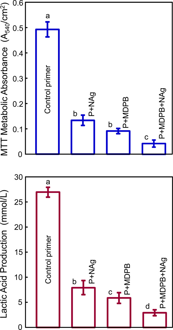
Results: Incorporating MDPB and NAg into primer did not reduce dentine bond strength compared to control (p>0.1). SEM revealed well-bonded adhesive-dentine interfaces with numerous resin tags. MDPB or NAg each greatly reduced biofilm viability and acid production, compared to control. Dual agents MDPB+NAg had a much stronger effect than either agent alone (p<0.05), increasing inhibition zone size and reducing metabolic activity, CFU and lactic acid by an order of magnitude, compared to control. There was no difference in cytotoxicity between commercial control and antibacterial primers (p>0.1).

Conclusions: The method of using dual agents MDPB+NAg in the primer yielded potent antibacterial properties. Hence, this method may be promising to combat residual bacteria in tooth cavity and invading bacteria at the margins. The dual agents MDPB+NAg may have wide applicability to other adhesives, composites, sealants and cements to inhibit biofilms and caries.


Figures

Figure 1
Figure 1
Un-cured primers in agar disk diffusion test. (A) Typical examples of inhibition zones of the four different primers against mutans streptococci. (B–D) Inhibition zone sizes for total microorganisms, total streptococci, and mutans streptococci, respectively. Each value is mean ± sd (n = 6). In each plot, dissimilar letters indicate values that are significantly different from each other (p < 0.05). NAg or MDPB alone each substantially increased the inhibition zone size. The use of dual agents MDPB and NAg together in the primer yielded the largest inhibition zones.
Figure 2
Figure 2
Bonding to dentin using extracted human molars: (A) Dentin shear bond strength (mean ± sd; n = 10), and (B) representative SEM image of the adhesive-dentin interfacial features. The four groups used different primers, but the same unmodified SBMP adhesive. In (A), all strength values were similar (p > 0.1). In (B), an example is shown for the control. All groups had similar dentin-adhesive interfacial features. Numerous resin tags were observed which were formed by the adhesive resin filling into the dentinal tubules.
Figure 3
Figure 3
Live/dead bacterial staining assay of biofilms adherent on the layered disks: (A) Schematic of biofilm experimental setup; (B–E) representative images of biofilms for control primer, P+NAg, P+MDPB, and P+MDPB+NAg. Live bacteria were stained green, and dead/compromised bacteria were stained red. Live and dead bacteria in close proximity of each other yielded yellow/orange colors.
Figure 4
Figure 4
MTT metabolic activity and lactic acid production of biofilms adherent on the layered disks (mean ± sd; n = 6). (A) MDPB or NAg alone each greatly decreased the metabolic activity. Dual agents (MDPB + NAg) yielded the lowest metabolic activity. (B) Lactic acid production of biofilms was substantially reduced by MDPB or NAg alone. The greatest reduction was achieved by using MDPB and NAg together in primer. In each plot, dissimilar letters indicate values that are significantly different from each other (p < 0.05).
Figure 5
Figure 5
Colony-forming unit counts (CFU) of biofilms adherent on the layered disks (mean ± sd; n = 6). (A) Total microorganisms, (B) total streptococci, and (C) mutans streptococci. In each plot, dissimilar letters indicate values that are significantly different from each other (p < 0.05). Adding MDPB or NAg into the primer imparted a potent antibacterial effect, and the strongest effect was achieved by combining MDPB and NAg in the same primer.
Figure 6
Figure 6
Human gingival fibroblast cytotoxicity (mean ± sd; n = 6). The culture medium consisted of fibroblast medium (FM) plus eluents from the cured primer/adhesive disks. Primer/adhesive volume (in unit mm3) divided by the total solution volume (mL) was 0.12, 0.24, and 0.47 mm3/mL, respectively. The 0.12 mm3/mL ratio corresponded to 6 disks in 1280 mL of solution, which compared to 1000 to 1500 mL/day of saliva for an average person. Even at higher concentrations of 0.24 and 0.47 mm3/mL, the resin eluent still had no adverse effect on the fibroblast viability. FM without resin eluents was used as control which had resin/solution volume ratio of 0. Fibroblast viability was measured by the MTT absorbance and normalized by FM control. All groups had cell viability matching that without resin eluents (p > 0.1).

Similar articles

Cited by

References

    1. Bagramian RA, Garcia-Godoy F, Volpe AR. The global increase in dental caries. A pending public health crisis. American Journal of Dentistry. 2009;22:3–8. - PubMed
    1. Dye BA, Tan S, Smith V, Lewis BG, Barker LK, Thornton-Evans G, et al. Trends in oral health status: United States, 1988–1994 and 1999–2004. Vital and Health Statistics. 2007;11:1–92. - PubMed
    1. Dye BA, Thornton-Evans G. Trends in oral health by poverty status as measured by Healthy People 2010 objectives. Public Health Reports. 2010;125:817–830. - PMC - PubMed
    1. Deng DM, ten Cate JM. Demineralization of dentin by Streptococcus mutans biofilms grown in the constant depth film fermentor. Caries Research. 2004;38:54–61. - PubMed
    1. Totiam P, Gonzalez-Cabezas C, Fontana MR, Zero DT. A new in vitro model to study the relationship of gap size and secondary caries. Caries Research. 2007;41:467–473. - PubMed

Publication types

MeSH terms