. 2013 Jun;7(3):543-54.
doi: 10.1016/j.molonc.2013.01.004. Epub 2013 Jan 19.

Suppression of the growth of human colorectal cancer cells by therapeutic stem cells expressing cytosine deaminase and interferon-β via their tumor-tropic effect in cellular and xenograft mouse models

Affiliations

Suppression of the growth of human colorectal cancer cells by therapeutic stem cells expressing cytosine deaminase and interferon-β via their tumor-tropic effect in cellular and xenograft mouse models

Bo-Rim Yi et al. Mol Oncol. 2013 Jun.

Abstract

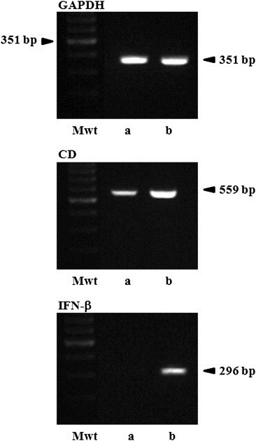
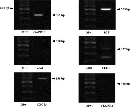
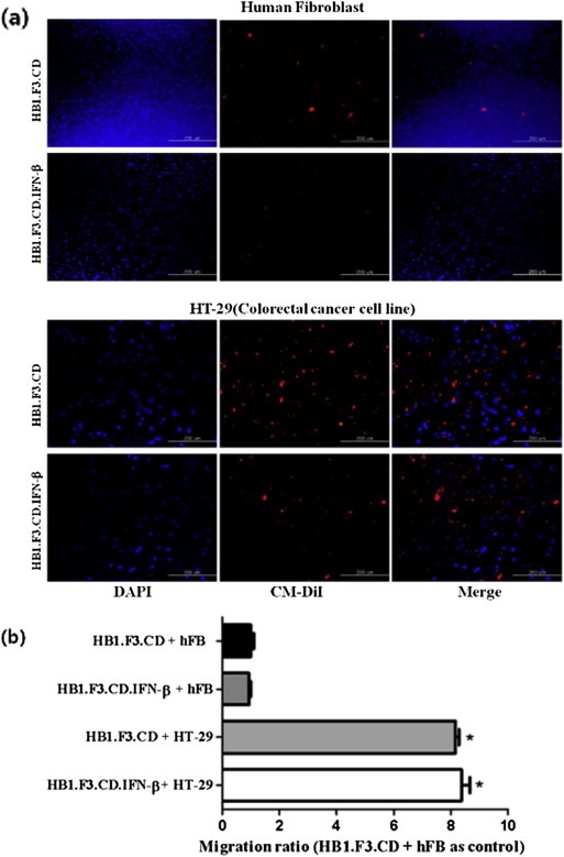
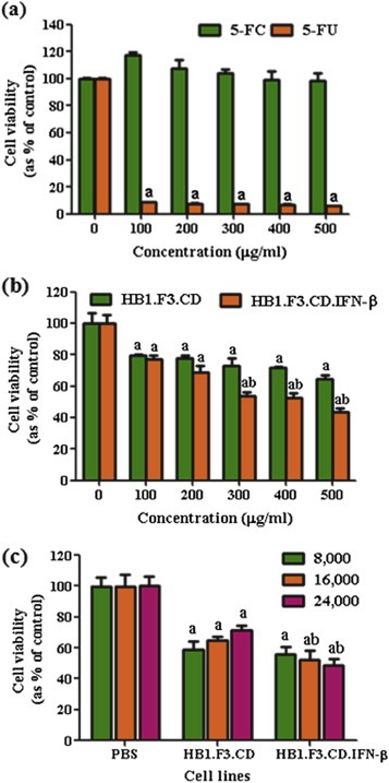
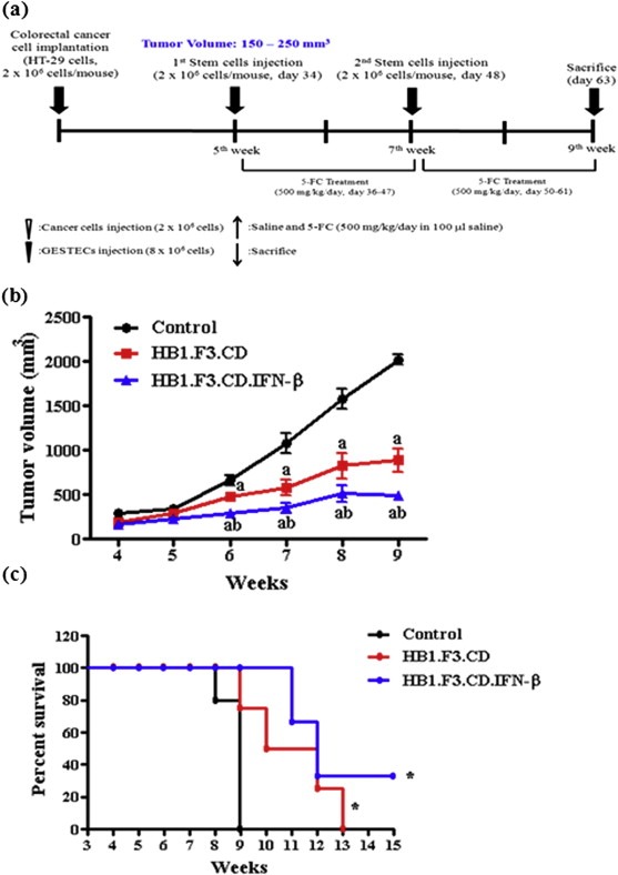
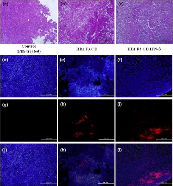
Genetically engineered stem cells (GESTECs) exhibit a potent therapeutic efficacy via their strong tumor tropism toward cancer cells. In this study, we introduced the human parental neural stem cells, HB1.F3, with the human interferon beta (IFN-β) gene which is a typical cytokine gene that has an antitumor effect and the cytosine deaminase (CD) gene from Escherichia coli (E. coli) that could convert the non-toxic prodrug, 5-fluorocytosine (5-FC), to a toxic metabolite, 5-fluorouracil (5-FU). Two types of stem cells expressing the CD gene (HB1.F3.CD cells) and both the CD and human IFN-β genes (HB1.F3.CD.IFN-β) were generated. The present study was performed to examine the migratory and therapeutic effects of these GESTECs against the colorectal cancer cell line, HT-29. When co-cultured with colorectal cancer cells in the presence of 5-FC, HB1.F3.CD and HB1.F3.CD.IFN-β cells exhibited the cytotoxicity on HT-29 cells via the bystander effect. In particular, HB1.F3.CD.IFN-β cells showed the synergistic cytotoxic activity of 5-FU and IFN-β. We also confirmed the migration ability of HB1.F3.CD and HB1.F3.CD.IFN-β cells toward HT-29 cells by a modified migration assay in vitro, where chemoattractant factors secreted by HT-29 cells attracted the GESTECs. In a xenograft mouse model, the volume of tumor mass was decreased up to 56% in HB1.F3.CD injected mice while the tumor mass was greatly inhibited about 76% in HB1.F3.CD.IFN-β injected mice. The therapeutic treatment by these GESTECs is a novel strategy where the combination of the migration capacity of stem cells as a vector for therapeutic genes towards colorectal cancer and a synergistic antitumor effect of CD and IFN-β genes can selectively target this type of cancer.


Figures

Figure 1
Figure 1
Expressions of E. coil CD and human IFN‐β genes in GESTECs. Expected products of E. coli CD or human IFN‐β genes in HB1.F3.CD and HB1.F3.CD.IFN‐β were shown at 559 bp and 296 bp, respectively. The cDNAs were synthesized from extracted RNAs of HB1.F3.CD and HB1.F3.CD.IFN‐β by RT and amplified by PCR. Next, the sizes of the PCR products were confirmed by 1.5% agarose gel electrophoresis. GAPDH was used as a control. a; HB1.F3.CD cells, b; HB1.F3.CD.IFN‐β cells, Mwt; molecular weight marker.
Figure 2
Figure 2
Expression of chemoattractant factors in colorectal cancer cells. After extracting total RNA from HT‐29 cells, RT‐PCR was performed. The expressions of chemoattractant ligands and receptors including SCF, VEGF, VEGFR2, CXCR4, and c‐kit, were identified by 1.5% agarose gel electrophoresis. Chemoattractant ligands and receptors, i.e., CXCR4, SCF, and VEGF, were highly expressed in HT‐29 cells. GAPDH was used an internal control Mwt: Molecular weight marker, NC: Negative control without cDNA template.
Figure 3
Figure 3
Migratory ability of GESTECs toward colorectal cancer cells. (a) Human fibroblast or HT‐29 (colorectal cancer cell; 1 × 105 cells/well) were seeded in the lower wells of 24‐well plates. HB1.F3.CD cells (1 × 105 cells/well) were stained with CD‐DiI and seeded in the fibronectin pre‐coated upper wells of 24‐well plates. DAPI staining solution was added to lower wells to observe colorectal cancer and human fibroblast. Blue stained cells indicated HT‐29 or human fibroblast as a control in the lower wells. Red stained cells indicated HB1.F3.CD or HB1.F3.CD.IFN‐β cells migrated from the upper wells toward HT‐29 colorectal or human fibroblast cells. (b) The migration ratio of stem cells was quantitated.
Figure 4
Figure 4
Effects of 5‐FC and 5‐FU on colorectal cancer cell growth. Proliferative rates at each concentration of 5‐FC or 5‐FU are expressed as relative fold‐change compared to the controls. (a) HT‐29 cells (4 × 103 cells/well) were seeded in 96‐well plates and treated with 5‐FC and 5‐FU at various concentrations (100, 200, 300, 400 or 500 μg/ml). (b) HB1.F3.CD and HB1.F3.CD.IFN‐β cells (8 × 103 cells/well) cultured with HT‐29 cells (4 × 103 cells/well) were treated with different concentrations of 5‐FC (100, 200, 300, 400, and 500 μg/ml). (c) HT‐29 cells (4 × 103 cells/well) were seeded in 96‐well plates and increasing numbers of HB1.F3.CD and HB1.F3.CD.IFN‐β cells (8 × 103, 1.6 × 104, or 2.4 × 104 cells/well) were added to the plates. The cells were then treated with 5‐FC or PBS (control) at a concentration of 500 μg/ml. Values are the mean ± SD for three independent experiments. a; P < 0.05 compared to the control. b; P < 0.05 compared the value of HB1.F3.CD.IFN‐β to HB1.F3.CD with identical number of cells.
Figure 5
Figure 5
Scheme of xenograft mouse model and the changes in tumor volume following GESTEC treatment. A xenograft model was employed implanting HT‐29 (2 × 106 cells) to male BALB/c nu/nu mice. (a) During 5 weeks, tumor mass volume mostly reached at 200 mm3. At day 34 and 48, pre‐stained with CM‐DiI human NSCs (4 × 106 cells/mouse) were injected surrounding xenografted tumor mass. After 2 days injected with GESTECs, 5‐FC (500 mg/kg/day) was treated every 24 h. At day 63, the mice were sacrificed. (b) The tumor volume of xenografted mice was measured caliper twice a week. Tumor volume was calculated by length × width × high × .5236. The volume of tumor mass was decreased up to 56% in HB1.F3.CD injected mice compared to a control. In addition, the tumor mass was inhibited about 74% in HB1.F3.CD.IFN‐β treated mice. (c) The Kaplan–Meier analysis and log rank test of mouse survival showed improved survival curve in the mice injected with GESTECs in the presence of 5‐FC compared to a control (P = 0.0437, by Log rank test for trend). a, P < 0.05 compared to the control; b, P < 0.05 compared the value of HB1.F3.CD only.
Figure 6
Figure 6
Histological analysis of xenografted tumor mass and fluorescent analysis of xenografted tumor mass. The xenografted tumor mass isolated at day 63 were fixed 10% neutral formalin and cut into 4–6 mm thick. Then, they were embedded in paraffin, sectioned 5 μm thick by a microtome. Tissue slides were stained with hematoxylin and eosin (H&E); (a)–(c). Injected GESTECs were pre‐labeled with CM‐DiI as cell tracker and prepared tissue slides were counterstained with DAPI solution. The stained tissue slides were observed under a microscope (Magnification: ×100, Blue: DAPI stained nuclei of colon cancer cells and GESTECs; (d), (e), (f). Red: CM‐DiI labeled GESTECs; (g), (h), (i). Merged; (j), (k), (l).). We observed red pre‐labeled stem cells surrounding or inside of tumor mass, indicating these GESTECs have a migratory ability in vivo. In addition less number of DAPI stained cells were observed at the surrounding areas located near red fluorescent cells.

Similar articles

Cited by

References

    1. Aboody, K.S. , Bush, R.A. , Garcia, E. , Metz, M.Z. , Najbauer, J. , Justus, K.A. , Phelps, D.A. , Remack, J.S. , Yoon, K.J. , Gillespie, S. , Kim, S.U. , Glackin, C.A. , Potter, P.M. , Danks, M.K. , 2006. Development of a tumor-selective approach to treat metastatic cancer. PLoS One 1, e23 - PMC - PubMed
    1. Aboody, K.S. , Najbauer, J. , Danks, M.K. , 2008. Stem and progenitor cell-mediated tumor selective gene therapy. Gene Ther. 15, 739–752. - PubMed
    1. Altaner, C. , 2008. Prodrug cancer gene therapy. Cancer Lett. 270, 191–201. - PubMed
    1. Bird, N.C. , Mangnall, D. , Majeed, A.W. , 2006. Biology of colorectal liver metastases: a review. J. Surg. Oncol. 94, 68–80. - PubMed
    1. Boucher, P.D. , Im, M.M. , Freytag, S.O. , Shewach, D.S. , 2006. A novel mechanism of synergistic cytotoxicity with 5-fluorocytosine and ganciclovir in double suicide gene therapy. Cancer Res. 66, 3230–3237. - PubMed

Publication types

MeSH terms