

Sources of variability of resting cerebral blood flow in healthy subjects: a study using ¹³³Xe SPECT measurements
- PMID: 23403374
- PMCID: PMC3652692
- DOI: 10.1038/jcbfm.2013.17
Sources of variability of resting cerebral blood flow in healthy subjects: a study using ¹³³Xe SPECT measurements
Abstract
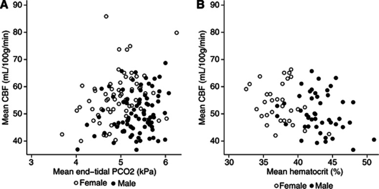
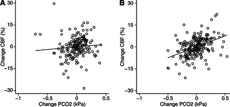
Measurements of cerebral blood flow (CBF) show large variability among healthy subjects. The aim of the present study was to investigate the relative effect of established factors influencing CBF on the variability of resting CBF. We retrospectively analyzed spontaneous variability in 430 CBF measurements acquired in 152 healthy, young subjects using (133)Xe single-photon emission computed tomography. Cerebral blood flow was correlated positively with both end-tidal expiratory PCO₂ (PETCO₂) and female gender and inversely with hematocrit (Hct). Between- and within-subject CO₂ reactivity was not significantly different. Including PETCO₂, Hct and gender in the model reduced between-subject and within-subject variance by 14% and 13.5%, respectively. Within-subject variability was mainly influenced by PETCO₂ and between-subject variability mostly by Hct, whereas gender appeared to be of little added value when Hct was also accounted for. The present study confirms large between-subject variability in CBF measurements and that gender, Hct, and PETCO₂ explain only a small part of this variability. This implies that a large fraction of CBF variability may be due to unknown factors such as differences in neuron density or metabolism that could be subject for further studies.
Figures
References
-
- Madsen PL, Holm S, Herning M, Lassen NA. Average blood flow and oxygen uptake in the human brain during resting wakefulness: a critical appraisal of the Kety-Schmidt technique. J Cereb Blood Flow Metab. 1993;13:646–655. - PubMed
-
- Ito H, Kanno I, Kato C, Sasaki T, Ishii K, Ouchi Y, et al. Database of normal human cerebral blood flow, cerebral blood volume, cerebral oxygen extraction fraction and cerebral metabolic rate of oxygen measured by positron emission tomography with 15O-labelled carbon dioxide or water, carbon monoxide and oxygen: a multicentre study in Japan. Eur J Nucl Med Mol Imaging. 2004;31:635–643. - PubMed
-
- Parkes LM, Rashid W, Chard DT, Tofts PS. Normal cerebral perfusion measurements using arterial spin labeling: reproducibility, stability, and age and gender effects. Magn Reson Med. 2004;51:736–743. - PubMed
-
- Henriksen OM, Larsson HB, Hansen AE, Gruner JM, Law I, Rostrup E. Estimation of intersubject variability of cerebral blood flow measurements using MRI and positron emission tomography. J Magn Reson Imaging. 2012;35:1290–1299. - PubMed
-
- Olesen J, Paulson OB, Lassen NA. Regional cerebral blood flow in man determined by the initial slope of the clearance of intra-arterially injected 133Xe. Stroke. 1971;2:519–540. - PubMed
Publication types
MeSH terms
Substances
LinkOut - more resources
Full Text Sources
Other Literature Sources
