

Long-term adaptation of cerebral hemodynamic response to somatosensory stimulation during chronic hypoxia in awake mice
- PMID: 23403375
- PMCID: PMC3652699
- DOI: 10.1038/jcbfm.2013.16
Long-term adaptation of cerebral hemodynamic response to somatosensory stimulation during chronic hypoxia in awake mice
Abstract
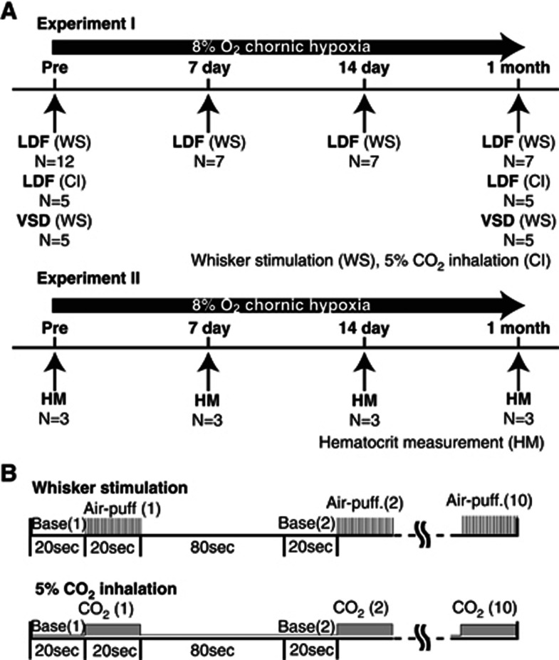
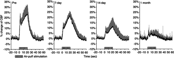
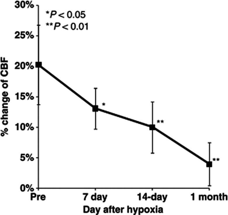
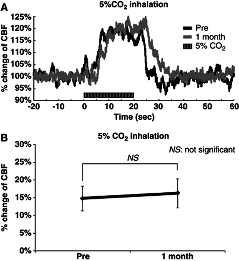
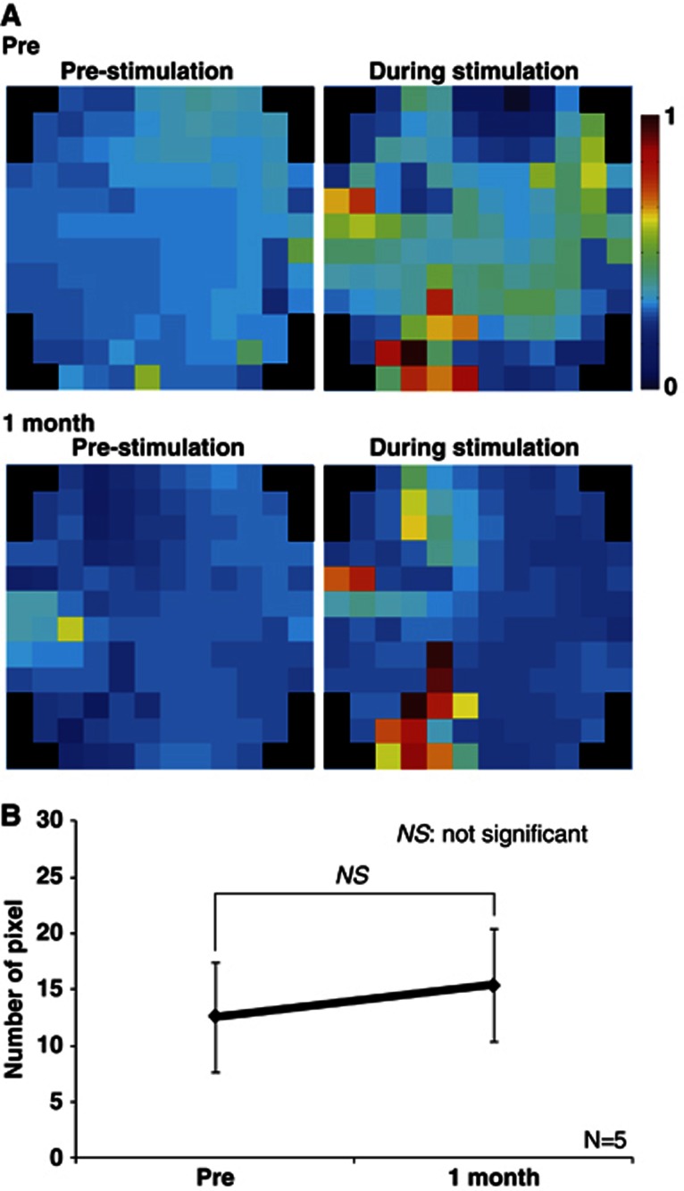
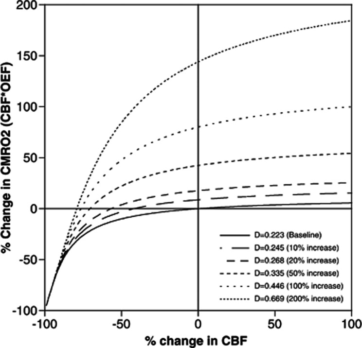
Effects of chronic hypoxia on hemodynamic response to sensory stimulation were investigated. Using laser-Doppler flowmetry, change in cerebral blood flow (CBF) was measured in awake mice, which were housed in a hypoxic chamber (8% O₂) for 1 month. The degree of increase in CBF evoked by sensory stimulation was gradually decreased over 1 month of chronic hypoxia. No significant reduction of increase in CBF induced by hypercapnia was observed during 1 month. Voltage-sensitive dye (VSD) imaging of the somatosensory cortex showed no significant decrease in neural activation over 1 month, indicating that the reduction of increase in CBF to sensory stimulation was not caused by cerebrovascular or neural dysfunction. The simulation study showed that, when effective diffusivity for oxygen in the capillary bed (D) value increases by chronic hypoxia due to an increase in capillary blood volume, an increase in the cerebral metabolic rate of oxygen utilization during neural activation can occur without any increase in CBF. Although previous study showed no direct effects of acute hypoxia on CBF response, our finding showed that hemodynamic response to neural activation could be modified in response to a change in their balance to energy demand using chronic hypoxia experiments.
Figures
Similar articles
-
Cerebral hemodynamic response to acute hyperoxia in awake mice.Brain Res. 2014 Apr 4;1557:155-63. doi: 10.1016/j.brainres.2014.01.053. Epub 2014 Feb 6. Brain Res. 2014. PMID: 24508909
-
Hemodynamic changes during somatosensory stimulation in awake and isoflurane-anesthetized mice measured by laser-Doppler flowmetry.Brain Res. 2012 Sep 7;1472:107-12. doi: 10.1016/j.brainres.2012.06.049. Epub 2012 Jul 10. Brain Res. 2012. PMID: 22789908
-
Cerebral blood flow adaptation to chronic hypoxia.Adv Exp Med Biol. 2008;614:371-7. doi: 10.1007/978-0-387-74911-2_41. Adv Exp Med Biol. 2008. PMID: 18290348
-
Control of cerebral blood flow during sleep and the effects of hypoxia.Adv Exp Med Biol. 2006;588:65-73. doi: 10.1007/978-0-387-34817-9_7. Adv Exp Med Biol. 2006. PMID: 17089880 Review.
-
Regulation of cerebral blood flow in mammals during chronic hypoxia: a matter of balance.Exp Physiol. 2010 Feb;95(2):251-62. doi: 10.1113/expphysiol.2008.045575. Epub 2009 Jul 17. Exp Physiol. 2010. PMID: 19617269 Review.
Cited by
-
Long-term effects of cerebral hypoperfusion on neural density and function using misery perfusion animal model.Sci Rep. 2016 Apr 27;6:25072. doi: 10.1038/srep25072. Sci Rep. 2016. PMID: 27116932 Free PMC article.
-
Changes in cortical microvasculature during misery perfusion measured by two-photon laser scanning microscopy.J Cereb Blood Flow Metab. 2014 Aug;34(8):1363-72. doi: 10.1038/jcbfm.2014.91. Epub 2014 May 21. J Cereb Blood Flow Metab. 2014. PMID: 24849667 Free PMC article.
-
Two-photon optogenetics-based assessment of neuronal connectivity in healthy and chronic hypoperfusion mice.Neurophotonics. 2024 Jul;11(3):035009. doi: 10.1117/1.NPh.11.3.035009. Epub 2024 Sep 28. Neurophotonics. 2024. PMID: 39345733 Free PMC article.
-
Developmental switch in neurovascular coupling in the immature rodent barrel cortex.PLoS One. 2013 Nov 5;8(11):e80749. doi: 10.1371/journal.pone.0080749. eCollection 2013. PLoS One. 2013. PMID: 24224059 Free PMC article.
-
Oxygen Enrichment Ameliorates Cardiorespiratory Alterations Induced by Chronic High-Altitude Hypoxia in Rats.Front Physiol. 2021 Jan 7;11:616145. doi: 10.3389/fphys.2020.616145. eCollection 2020. Front Physiol. 2021. PMID: 33488404 Free PMC article.
References
-
- LaManna JC, Chavez JC, Pichiule P. Structural and functional adaptation to hypoxia in the rat brain. J Exp Biol. 2004;207:3163–3169. - PubMed
-
- Ainslie PN, Ogoh S. Regulation of cerebral blood flow in mammals during chronic hypoxia: a matter of balance. Exp Physiol. 2010;95:251–262. - PubMed
-
- Wilson M, Newman HS, Imray C. The cerebral effects of ascent to high altitudes. Lancet Neurol. 2009;8:175–191. - PubMed
-
- Jensen JB, Sperling B, Severinghaus JW, Lassen NA. Augmented hypoxic cerebral vasodilation in men during 5 days at 3,810 m altitude. J Appl Physiol. 1996;80:1214–1218. - PubMed
-
- Pichiule P, LaManna JC. Angiopoietin-2 and rat brain capillary remodeling during adaptation and deadaptation to prolonged mild hypoxia. J Appl Physiol. 2002;93:1131–1139. - PubMed
MeSH terms
Substances
LinkOut - more resources
Full Text Sources
Other Literature Sources
