Identification of Francisella novicida mutants that fail to induce prostaglandin E(2) synthesis by infected macrophages
- PMID: 23403609
- PMCID: PMC3568750
- DOI: 10.3389/fmicb.2013.00016
Identification of Francisella novicida mutants that fail to induce prostaglandin E(2) synthesis by infected macrophages
Abstract
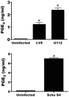
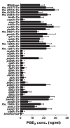
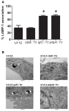
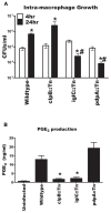
Francisella tularensis is the causative agent of tularemia. We have previously shown that infection with F. tularensis Live Vaccine Strain (LVS) induces macrophages to synthesize prostaglandin E(2) (PGE(2)). Synthesis of PGE(2) by F. tularensis infected macrophages results in decreased T cell proliferation in vitro and increased bacterial survival in vivo. Although we understand some of the biological consequences of F. tularensis induced PGE(2) synthesis by macrophages, we do not understand the cellular pathways (neither host nor bacterial) that result in up-regulation of the PGE(2) biosynthetic pathway in F. tularensis infected macrophages. We took a genetic approach to begin to understand the molecular mechanisms of bacterial induction of PGE(2) synthesis from infected macrophages. To identify F. tularensis genes necessary for the induction of PGE(2) in primary macrophages, we infected cells with individual mutants from the closely related strain F. tularensis subspecies novicida U112 (U112) two allele mutant library. Twenty genes were identified that when disrupted resulted in U112 mutant strains unable to induce the synthesis of PGE(2) by infected macrophages. Fourteen of the genes identified are located within the Francisella pathogenicity island (FPI). Genes in the FPI are required for F. tularensis to escape from the phagosome and replicate in the cytosol, which might account for the failure of U112 with transposon insertions within the FPI to induce PGE(2). This implies that U112 mutant strains that do not grow intracellularly would also not induce PGE(2). We found that U112 clpB::Tn grows within macrophages yet fails to induce PGE(2), while U112 pdpA::Tn does not grow yet does induce PGE(2). We also found that U112 iglC::Tn neither grows nor induces PGE(2). These findings indicate that there is dissociation between intracellular growth and the ability of F. tularensis to induce PGE(2) synthesis. These mutants provide a critical entrée into the pathways used in the host for PGE(2) induction.
Keywords: Francisella; prostaglandin E.
Figures


Similar articles
-
Perforin- and granzyme-mediated cytotoxic effector functions are essential for protection against Francisella tularensis following vaccination by the defined F. tularensis subsp. novicida ΔfopC vaccine strain.Infect Immun. 2012 Jun;80(6):2177-85. doi: 10.1128/IAI.00036-12. Epub 2012 Apr 9. Infect Immun. 2012. PMID: 22493083 Free PMC article.
-
The Francisella tularensis pathogenicity island protein IglC and its regulator MglA are essential for modulating phagosome biogenesis and subsequent bacterial escape into the cytoplasm.Cell Microbiol. 2005 Jul;7(7):969-79. doi: 10.1111/j.1462-5822.2005.00526.x. Cell Microbiol. 2005. PMID: 15953029
-
Mutations of Francisella novicida that alter the mechanism of its phagocytosis by murine macrophages.PLoS One. 2010 Jul 29;5(7):e11857. doi: 10.1371/journal.pone.0011857. PLoS One. 2010. PMID: 20686600 Free PMC article.
-
Effects of the putative transcriptional regulator IclR on Francisella tularensis pathogenesis.Infect Immun. 2010 Dec;78(12):5022-32. doi: 10.1128/IAI.00544-10. Epub 2010 Oct 4. Infect Immun. 2010. PMID: 20921148 Free PMC article.
-
Molecular and genetic basis of pathogenesis in Francisella tularensis.Ann N Y Acad Sci. 2007 Jun;1105:138-59. doi: 10.1196/annals.1409.010. Epub 2007 Mar 29. Ann N Y Acad Sci. 2007. PMID: 17395737 Review.
Cited by
-
Adaptive Immunity to Francisella tularensis and Considerations for Vaccine Development.Front Cell Infect Microbiol. 2018 Apr 6;8:115. doi: 10.3389/fcimb.2018.00115. eCollection 2018. Front Cell Infect Microbiol. 2018. PMID: 29682484 Free PMC article. Review.
-
Janus kinase 3 activity is necessary for phosphorylation of cytosolic phospholipase A2 and prostaglandin E2 synthesis by macrophages infected with Francisella tularensis live vaccine strain.Infect Immun. 2014 Mar;82(3):970-82. doi: 10.1128/IAI.01461-13. Epub 2013 Dec 16. Infect Immun. 2014. PMID: 24343645 Free PMC article.
-
Host-pathogen interactions and immune evasion strategies in Francisella tularensis pathogenicity.Infect Drug Resist. 2014 Sep 18;7:239-51. doi: 10.2147/IDR.S53700. eCollection 2014. Infect Drug Resist. 2014. PMID: 25258544 Free PMC article. Review.
-
Francisella tularensis LVS induction of prostaglandin biosynthesis by infected macrophages requires specific host phospholipases and lipid phosphatases.Infect Immun. 2014 Aug;82(8):3299-311. doi: 10.1128/IAI.02060-14. Epub 2014 May 27. Infect Immun. 2014. PMID: 24866789 Free PMC article.
-
Lipin-1 Contributes to IL-4 Mediated Macrophage Polarization.Front Immunol. 2020 May 5;11:787. doi: 10.3389/fimmu.2020.00787. eCollection 2020. Front Immunol. 2020. PMID: 32431707 Free PMC article.
References
-
- Allison L. A., Moyle M., Shales M., Ingles C. J. (1985). Extensive homology among the largest subunits of eukaryotic and prokaryotic RNA polymerases. Cell 42 599–610 - PubMed
Grants and funding
LinkOut - more resources
Full Text Sources
Other Literature Sources

