. 2013 Apr 11;121(15):2943-51.
doi: 10.1182/blood-2012-10-463166. Epub 2013 Feb 12.

The risk of hemophagocytic lymphohistiocytosis in Hermansky-Pudlak syndrome type 2

Affiliations

The risk of hemophagocytic lymphohistiocytosis in Hermansky-Pudlak syndrome type 2

Birthe Jessen et al. Blood. .

Abstract

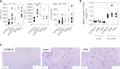
Genetic disorders of lymphocyte cytotoxicity predispose patients to hemophagocytic lymphohistiocytosis (HLH). Reduced lymphocyte cytotoxicity has been demonstrated in Hermansky-Pudlak syndrome type 2 (HPS2), but only a single patient was reported who developed HLH. Because that patient also carried a potentially contributing heterozygous RAB27A mutation, the risk for HLH in HPS2 remains unclear. We analyzed susceptibility to HLH in the pearl mouse model of HPS2. After infection with lymphocytic choriomeningitis virus, pearl mice developed all key features of HLH, linked to impaired virus control caused by a moderate defect in CTL cytotoxicity in vivo. However, in contrast to perforin-deficient mice, the disease was transient, and all mice fully recovered and controlled the infection. An additional heterozygous Rab27a mutation did not aggravate the cytotoxicity defect or disease parameters. In the largest survey of 22 HPS2 patients covering 234 patient years, we identified only 1 additional patient with HLH and 2 with incomplete transient HLH-like episodes, although cytotoxicity or degranulation was impaired in all 16 patients tested. HPS2 confers a risk for HLH that is lower than in Griscelli or Chediak-Higashi syndrome, probably because of a milder defect in cytotoxicity. Preemptive hematopoietic stem cell transplantation does not appear justified in HPS2.


Figures

Figure 1
Figure 1
Pearl mice develop transient HLH after LCMV infection independent of the presence of an additional Rab27a mutation. C57BL/6, pearl, pearl-ash+/−, ashen, and PKO mice were infected intravenously with 200 pfu of LCMV-WE, and (A) body weight and (B) temperature were monitored daily. Dashed lines indicate the detection limit. (C) Blood counts were analyzed on d8 after infection. The white line indicates the mean, and the gray area indicates the range of values of naïve pearl mice. D8 serum levels of (D) ferritin, (E) sCD25, and (F) IFN-γ are shown. Graphs show pooled data of 2 independent experiments with 3-4 mice per group except for ashen mice, in which results from 3 mice obtained in 2 independent experiments are shown. n.s. indicates not significant (P > .05); *P < .05; **P < .01; ***P < .001.
Figure 2
Figure 2
Liver disease and histopathological alterations in pearl mice undergoing LCMV-induced HLH. C57BL/6, pearl, pearl-ash+/−, ashen, and PKO mice were infected with LCMV-WE and 8 days later, serum levels of (A) GPT, LDH, and triglycerides (TG) were determined. (B) Splenomegaly was analyzed by calculating spleen weight in percent of body weight. (C) Representative liver sections stained with anti-F4/80 (original magnification, 10×) obtained at day 8 after infection. Lower left inset: semiquantitative analysis of hemophagocyting macrophages as assessed in 10 high-power visual fields (40×) per mouse. (+) indicates rare; +, few; and ++, frequent. Scale bars represent 100 µm. n.s. indicates not significant (P > .05); *P < .05; **P < .01.
Figure 3
Figure 3
Impaired NK-cell function in pearl mice. C57BL/6, pearl, and PKO mice were injected intraperitoneally with 200 µg of poly (I:C). At 24 hours later, spleen cells were restimulated for 2 hours with YAC-1 target cells or medium as a control in the presence of anti-CD107a antibody (A-B). (A) Representative histograms of NK1.1+CD3 NK cells are shown. Dashed line indicates medium control; solid line indicates restimulation with YAC-1 cells. (B) Degranulation is shown as increase of CD107a expression on NK1.1+CD3- cells (ΔCD107a) after restimulation with YAC-1 cells compared with medium control. (C) NK-cell cytotoxicity was determined in a 5-hour 51chromium release assay on YAC-1 target cells. Quantification of NK cells was performed by flow cytometry. Representative data for 3 independent experiments with 3-5 mice/group are shown.
Figure 4
Figure 4
Pearl mice show a delay in virus control. C57BL/6, pearl, pearl-ash+/−, ashen, and PKO mice were infected with 200 pfu of LCMV-WE and (A) virus titer in spleens were determined on days 4, 8, and 12 after infection. (B) Days 8 and 12 virus titers are shown for lung and liver. The means and the SDs are indicated. Graphs show pooled data of 2 independent experiments with 3-4 mice per group. For ashen mice data of 3 mice obtained in 2 different experiments are shown. The dashed lines indicate the detection limit.
Figure 5
Figure 5
CTL of pearl mice are impaired in degranulation and cytotoxicity. CTL from C57BL/6, pearl, ashen, and PKO mice were analyzed 8 days after infection with 200 pfu of LCMV-WE. Degranulation of LCMV-specific CTL was analyzed after in vitro restimulation with the gp33 peptide. (A) Representative fluorescence-activated cell-sorting plots gated on CD3+CD8+ lymphocytes are shown. Numbers indicate the percentage of CD107a+ cells among gp33-specific IFN-γ+ CTL. (B) Frequencies of CD107a+ cells of IFN-γ+CD8+ T cells were analyzed. Pooled data from 3 independent experiments are shown. ***P < .001 (C) Wild-type mice were infected with 104 pfu LCMV. Then, 10 hours later they were adoptively transfused with 2 × 10⁶ isolated day 8 splenic CTL of either wild-type or pearl mice and after additional 18-hour splenic virus titers were analyzed. Pooled data from 2 independent experiments with 3-4 mice per group are shown. The dashed line indicates the detection limit. Nil indicates without transfer. (D) Ex vivo cytotoxicity was tested in a 5-hour 51chromiumrelease assay on either LCMV-infected MC57 target cells (left) or gp33 peptide-loaded EL-4 target cells (right). Results from 1 of 2 independent experiments with 3 mice per group are shown.
Figure 6
Figure 6
Function and phenotype of pearl CTL at different time points after infection. (A, B) Cytokine production of C57BL/6, pearl, and PKO CTL was analyzed by flow cytometry on days 8 and 12 after infection with 200 pfu of LCMV. CTL were stained for (A) IFN-γ and (B) TNF-α production after peptide stimulation in vitro. Representative FACS plots are shown at the top and (A) frequencies of IFN-γhi cells among IFN-γ+ CD8 T cells and (B) frequencies of TNF-α+ cells among CD8 T cells are shown at the bottom. Surface expression of (C) KLRG1 and CD127 and (D) CD44 and CD62L on CTL are depicted as representative FACS plots (top) and frequencies are shown (bottom). Representative data from 1 of 2 independent experiments with 3-4 mice per group are shown. For IFN-γ and TNF-α (day 8) pooled data from two independent experiments are shown. n.s. indicates not significant (P > .05); *P < .05; **P < .01; ***P < .001.

Similar articles

Cited by

  • Hermansky-Pudlak syndrome type 2 manifests with fibrosing lung disease early in childhood.
    Hengst M, Naehrlich L, Mahavadi P, Grosse-Onnebrink J, Terheggen-Lagro S, Skanke LH, Schuch LA, Brasch F, Guenther A, Reu S, Ley-Zaporozhan J, Griese M. Hengst M, et al. Orphanet J Rare Dis. 2018 Mar 27;13(1):42. doi: 10.1186/s13023-018-0780-z. Orphanet J Rare Dis. 2018. PMID: 29580292 Free PMC article.
  • A Novel Likely Pathogenic Variant in the BLOC1S5 Gene Associated with Hermansky-Pudlak Syndrome Type 11 and an Overview of Human BLOC-1 Deficiencies.
    Boeckelmann D, Wolter M, Käsmann-Kellner B, Koehler U, Schieber-Nakamura L, Zieger B. Boeckelmann D, et al. Cells. 2021 Oct 1;10(10):2630. doi: 10.3390/cells10102630. Cells. 2021. PMID: 34685610 Free PMC article. Review.
  • A homozygous AP3D1 missense variant in patients with sensorineural hearing loss as the leading manifestation.
    Frohne A, Koenighofer M, Cetin H, Nieratschker M, Liu DT, Laccone F, Neesen J, Nemec SF, Schwarz-Nemec U, Schoefer C, Avraham KB, Frei K, Grabmeier-Pfistershammer K, Kratzer B, Schmetterer K, Pickl WF, Parzefall T. Frohne A, et al. Hum Genet. 2023 Aug;142(8):1077-1089. doi: 10.1007/s00439-022-02506-0. Epub 2022 Nov 29. Hum Genet. 2023. PMID: 36445457 Free PMC article.
  • Too much of a good thing: a review of primary immune regulatory disorders.
    Tsilifis C, Slatter MA, Gennery AR. Tsilifis C, et al. Front Immunol. 2023 Oct 31;14:1279201. doi: 10.3389/fimmu.2023.1279201. eCollection 2023. Front Immunol. 2023. PMID: 38022498 Free PMC article. Review.
  • Fourth Update on the Iranian National Registry of Primary Immunodeficiencies: Integration of Molecular Diagnosis.
    Abolhassani H, Kiaee F, Tavakol M, Chavoshzadeh Z, Mahdaviani SA, Momen T, Yazdani R, Azizi G, Habibi S, Gharagozlou M, Movahedi M, Hamidieh AA, Behniafard N, Nabavi M, Bemanian MH, Arshi S, Molatefi R, Sherkat R, Shirkani A, Amin R, Aleyasin S, Faridhosseini R, Jabbari-Azad F, Mohammadzadeh I, Ghaffari J, Shafiei A, Kalantari A, Mansouri M, Mesdaghi M, Babaie D, Ahanchian H, Khoshkhui M, Soheili H, Eslamian MH, Cheraghi T, Dabbaghzadeh A, Tavassoli M, Kalmarzi RN, Mortazavi SH, Kashef S, Esmaeilzadeh H, Tafaroji J, Khalili A, Zandieh F, Sadeghi-Shabestari M, Darougar S, Behmanesh F, Akbari H, Zandkarimi M, Abolnezhadian F, Fayezi A, Moghtaderi M, Ahmadiafshar A, Shakerian B, Sajedi V, Taghvaei B, Safari M, Heidarzadeh M, Ghalebaghi B, Fathi SM, Darabi B, Bazregari S, Bazargan N, Fallahpour M, Khayatzadeh A, Javahertrash N, Bashardoust B, Zamani M, Mohsenzadeh A, Ebrahimi S, Sharafian S, Vosughimotlagh A, Tafakoridelbari M, Rahimi M, Ashournia P, Razaghian A, Rezaei A, Mamishi S, Parvaneh N, Rezaei N, Hammarström L, Aghamohammadi A. Abolhassani H, et al. J Clin Immunol. 2018 Oct;38(7):816-832. doi: 10.1007/s10875-018-0556-1. Epub 2018 Oct 9. J Clin Immunol. 2018. PMID: 30302726

References

    1. Hermansky F, Pudlak P. Albinism associated with hemorrhagic diathesis and unusual pigmented reticular cells in the bone marrow: report of two cases with histochemical studies. Blood. 1959;14(2):162–169. - PubMed
    1. Shotelersuk V, Dell’Angelica EC, Hartnell L, et al. A new variant of Hermansky-Pudlak syndrome due to mutations in a gene responsible for vesicle formation. Am J Med. 2000;108(5):423–427. - PubMed
    1. Badolato R, Parolini S. Novel insights from adaptor protein 3 complex deficiency. J Allergy Clin Immunol. 2007;120(4):735–741. quiz 742-733. - PubMed
    1. Clark R, Griffiths GM. Lytic granules, secretory lysosomes and disease. Curr Opin Immunol. 2003;15(5):516–521. - PubMed
    1. Enders A, Zieger B, Schwarz K, et al. Lethal hemophagocytic lymphohistiocytosis in Hermansky-Pudlak syndrome type II. Blood. 2006;108(1):81–87. - PubMed

Publication types

MeSH terms