

doi: 10.1117/1.JBO.18.2.026020.
Color-matched and fluorescence-labeled esophagus phantom and its applications
Affiliations
- PMID: 23403908
- PMCID: PMC3569733
- DOI: 10.1117/1.JBO.18.2.026020
Item in Clipboard
Color-matched and fluorescence-labeled esophagus phantom and its applications
J Biomed Opt.
2013 Feb.
Abstract
We developed a stable, reproducible three-dimensional optical phantom for the evaluation of a wide-field endoscopic molecular imaging system. This phantom mimicked a human esophagus structure with flexibility to demonstrate body movements. At the same time, realistic visual appearance and diffuse spectral reflectance properties of the tissue were simulated by a color matching methodology. A photostable dye-in-polymer technology was applied to represent biomarker probed "hot-spot" locations. Furthermore, fluorescent target quantification of the phantom was demonstrated using a 1.2 mm ultrathin scanning fiber endoscope with concurrent fluorescence-reflectance imaging.
Figures
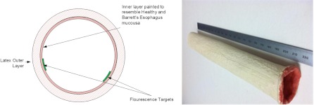
(a) A cross-sectional graph illustrating the phantom design. (b) An overview of the resultant phantom.
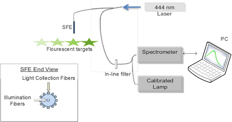
Schematic diagram of the experimental setup to quantify targets’ fluorescence.
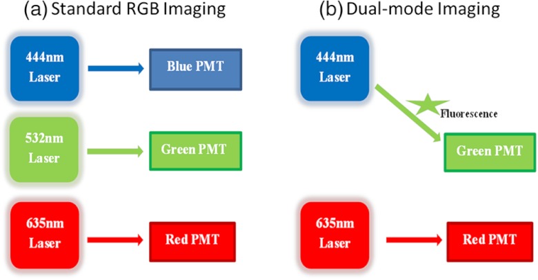
(a) Standard SFE RGB imaging; (b) SFE dual mode imaging, with the 532 nm blue photomultiplier tube (PMT) channel inactive.
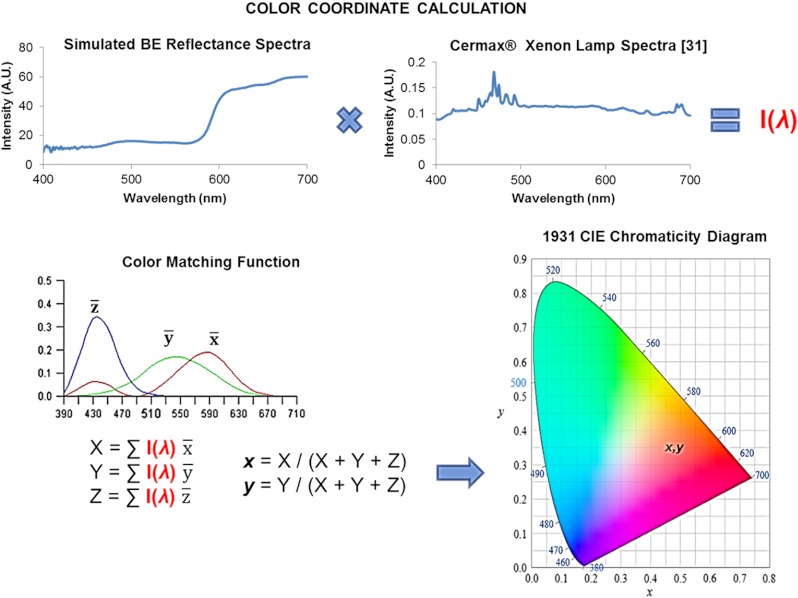
A summary diagram of the CIE color calculation methodology. Calculated color coordinates represent the visual appearance of a paint recipe viewed under illumination by the xenon Cermax lamp. Clinically BE is observed with an endoscope that incorporates a color CCD camera. The spectral response of modern endoscopic CCD cameras closely matches the ideal , , and functions. Therefore, the calculated color coordinates correspond reasonably well to the clinically observed color of BE.
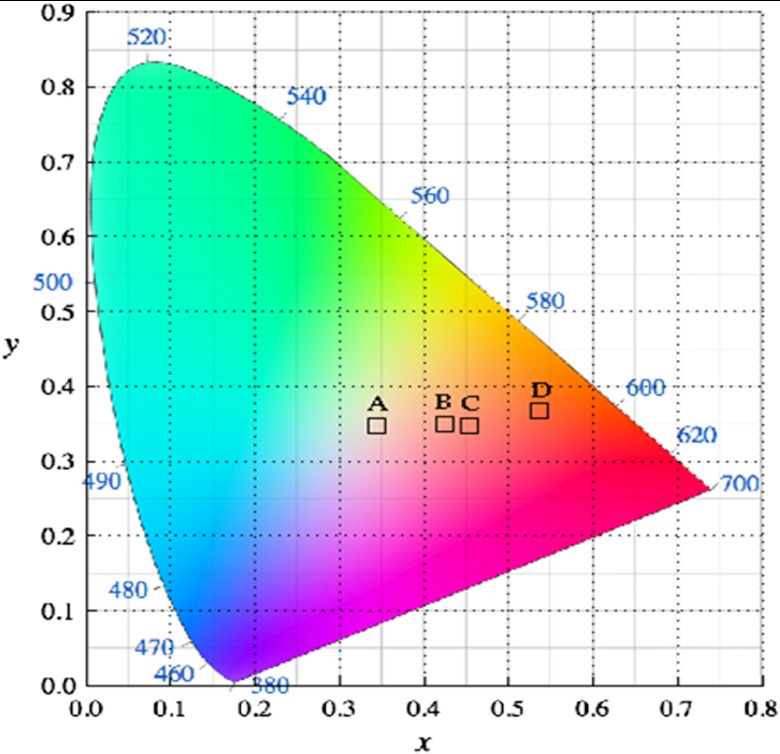
CIE color calculations of: (a) Ref. BE color, (b) simulated healthy esophagus mucosa color, (c) simulated BE color, and (d) Atlantic salmon fillet color.
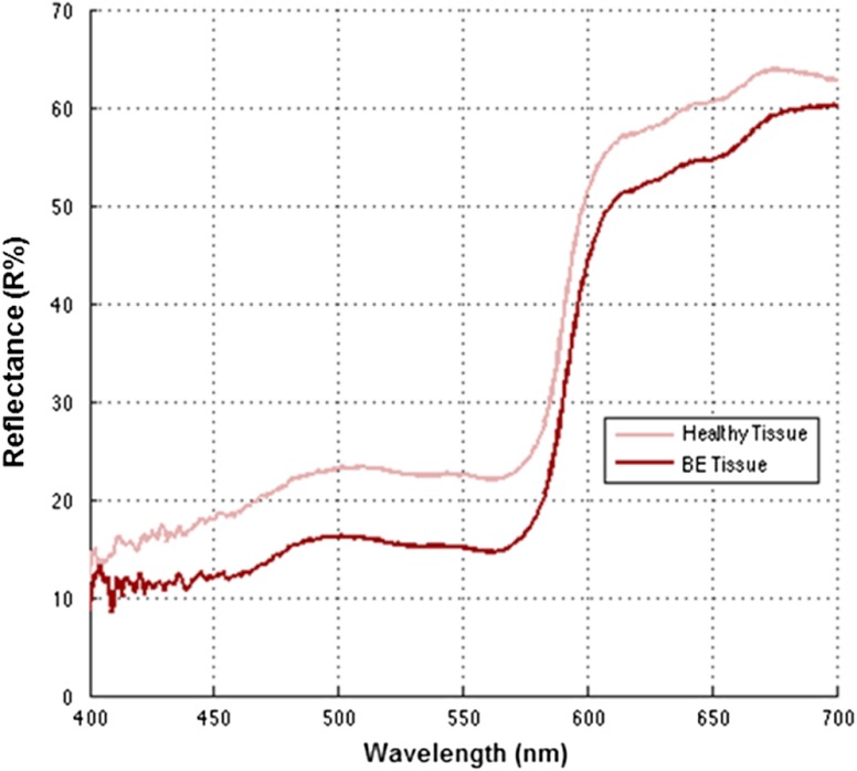
Diffuse spectra reflectance of simulated healthy esophagus and BE tissue from the phantom.
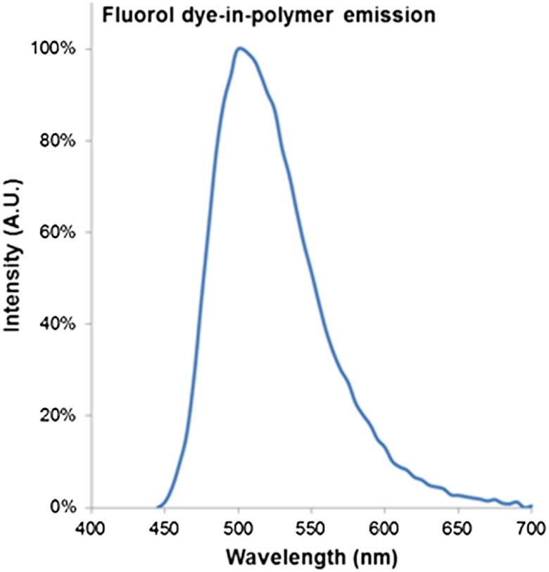
Fluorol dye-in-polymer emission spectra under 444 nm laser excitation, measured by a calibrated spectrometer.
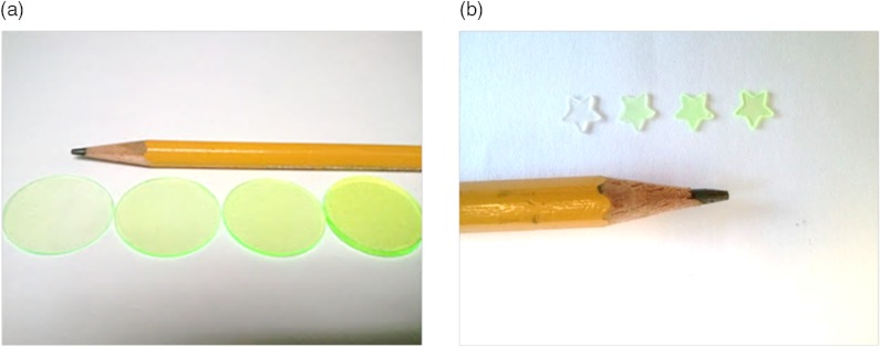
Photo representations of dye-in-polymer. (a) thin disks, (b) die-cut distinctive star shaped targets.
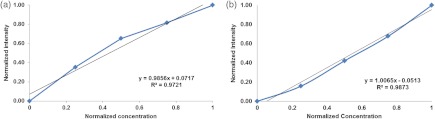
(a) Fluorescent target emission intensity recorded with a spectrometer as a function of dye concentration. (b) Fluorescent target SFE image intensity as a function of dye concentration.
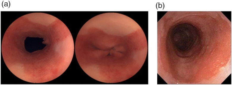
Standard RGB SFE imaging of the phantom. (a) SFE images of the same phantom with with sphincter open (left) and sphincter closed (right). (b) Endoscope images of a human Barrett’s esophagus © 2004 by Mayo Foundation for Medical Education and Research.
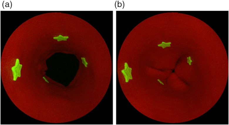
SFE fluorescence and red reflectance dual-modal imaging of the phantom. Dual-modal SFE imaging of the phantom with four fluorescent targets. Left: sphincter open. Right: sphincter closed.
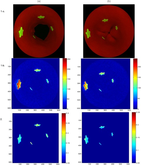
Phantom application: the SFE distance compensation (DC) algorithm development. (a) Sphincter open mode; (b) sphincter closed mode; (1-a) before DC SFE dual-mode (reflectance-fluorescence) image; (1-b) before DC colormap of the fluorescence image from (1-a); (2) After DC colormap of the fluorescence image from (1-a).
Similar articles
-
Target-to-background enhancement in multispectral endoscopy with background autofluorescence mitigation for quantitative molecular imaging.J Biomed Opt. 2014;19(7):76014. doi: 10.1117/1.JBO.19.7.076014. J Biomed Opt. 2014. PMID: 25027002 Free PMC article.
-
Automated coregistered imaging using a hand-held probe-based optical imager.Rev Sci Instrum. 2010 Feb;81(2):023702. doi: 10.1063/1.3271019. Rev Sci Instrum. 2010. PMID: 20192497
-
Comprehensive phantom for interventional fluorescence molecular imaging.J Biomed Opt. 2016 Sep;21(9):091309. doi: 10.1117/1.JBO.21.9.091309. J Biomed Opt. 2016. PMID: 27304578
-
The combined use of fluorescence, reflectance, and light-scattering spectroscopy for evaluating dysplasia in Barrett's esophagus.Gastrointest Endosc Clin N Am. 2004 Jul;14(3):519-37, ix. doi: 10.1016/j.giec.2004.03.008. Gastrointest Endosc Clin N Am. 2004. PMID: 15261200 Review.
-
Characterization of dysplastic tissue morphology and biochemistry in Barrett's esophagus using diffuse reflectance and light scattering spectroscopy.Gastrointest Endosc Clin N Am. 2003 Apr;13(2):297-308. doi: 10.1016/s1052-5157(03)00008-4. Gastrointest Endosc Clin N Am. 2003. PMID: 12916661 Review.
Cited by
-
Target-to-background enhancement in multispectral endoscopy with background autofluorescence mitigation for quantitative molecular imaging.J Biomed Opt. 2014;19(7):76014. doi: 10.1117/1.JBO.19.7.076014. J Biomed Opt. 2014. PMID: 25027002 Free PMC article.
-
Standardization and implementation of fluorescence molecular endoscopy in the clinic.J Biomed Opt. 2022 Feb;27(7):074704. doi: 10.1117/1.JBO.27.7.074704. J Biomed Opt. 2022. PMID: 35170264 Free PMC article.
-
Mapping surgical fields by moving a laser-scanning multimodal scope attached to a robot arm.Proc SPIE Int Soc Opt Eng. 2014 Feb;9036:90362S. doi: 10.1117/12.2044165. Epub 2014 Mar 12. Proc SPIE Int Soc Opt Eng. 2014. PMID: 34321710 Free PMC article.
-
Multimodal laser-based angioscopy for structural, chemical and biological imaging of atherosclerosis.Nat Biomed Eng. 2017;1:0023. doi: 10.1038/s41551-016-0023. Epub 2017 Feb 10. Nat Biomed Eng. 2017. PMID: 28555172 Free PMC article.
-
Toward real-time quantification of fluorescence molecular probes using target/background ratio for guiding biopsy and endoscopic therapy of esophageal neoplasia.J Med Imaging (Bellingham). 2017 Apr;4(2):024502. doi: 10.1117/1.JMI.4.2.024502. Epub 2017 May 24. J Med Imaging (Bellingham). 2017. PMID: 28560244 Free PMC article.
References
Publication types
MeSH terms
Substances
Grants and funding
LinkOut - more resources
Full Text Sources
Other Literature Sources
