. 2013 Mar 20;32(6):886-98.
doi: 10.1038/emboj.2013.23. Epub 2013 Feb 12.

Signal recognition initiates reorganization of the presequence translocase during protein import

Affiliations

Signal recognition initiates reorganization of the presequence translocase during protein import

Oleksandr Lytovchenko et al. EMBO J. .

Abstract

The mitochondrial presequence translocase interacts with presequence-containing precursors at the intermembrane space (IMS) side of the inner membrane to mediate their translocation into the matrix. Little is known as too how these matrix-targeting signals activate the translocase in order to initiate precursor transport. Therefore, we analysed how signal recognition by the presequence translocase initiates reorganization among Tim-proteins during import. Our analyses revealed that the presequence receptor Tim50 interacts with Tim21 in a signal-sensitive manner in a process that involves the IMS-domain of the Tim23 channel. The signal-driven release of Tim21 from Tim50 promotes recruitment of Pam17 and thus triggers formation of the motor-associated form of the TIM23 complex required for matrix transport.


Conflict of interest statement

The authors declare that they have no conflict of interest.

Figures

Figure 1
Figure 1
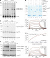
Identification of Tim50 crosslinking partners. (A) Schematic representation of Tim50 and Tim50 fragments used in this study. Presequence (PS), transmembrane domain (TM), NIF-domain, presequence-binding domain (PBD), and the cysteine position (C268) are indicated. (B) Structure of Tim50C (Protein Data Bank entry 3QLE) with the Tim23-binding site, large negatively charged groove and the cysteine (C268, in green) positions indicated. Positively charged residues are coloured in blue, negative are red. (C) Wild-type and Tim50HA-mitochondria were subjected to CuSO4 crosslinking and analysed by western blotting using either anti-Tim50 or anti-HA antibodies. Selected shifted crosslinking products are marked with circles or a diamond (unknown Tim50 crosslinking partner). (D) Crosslinking was performed as in (C) using Tim21ProtA-, Tim23ProtA- and Tim23ProtA-mitochondria. Samples were analysed by western blotting using anti-Tim50 antiserum. (E) CuSO4 crosslinking was performed in mitochondria from Tim21ProtA and tim21Δ strains, transformed with either an empty pFL39 (control) or a Tim21-expressing plasmid (TIM21). Samples were analysed by western blotting using an antiserum against Tim50.
Figure 2
Figure 2
Tim21 interacts directly with Tim50 in organello and in vitro. (A) Wild-type mitochondria were osmotically swollen and subjected to CuSO4 crosslinking in the presence of indicated concentrations of purified Tim21IMS or its His-tagged form Tim21His. Crosslinking-adducts were detected by immunoblotting against Tim50. (B) Purified recombinant proteins (25 μM Tim21IMS or Tim21C128S and 10 μM Tim50IMS) were crosslinked in the presence of 5 mM CuSO4. Crosslinking-adducts were visualized by colloidal Coomassie staining. (C) Indicated amounts of purified Tim50IMS or bovine serum albumin (BSA) were mixed with recombinant Tim21IMS (final concentration 1 μM) and crosslinked by adding of CuSO4, followed by immunoblotting and decoration with anti-Tim21 antibody. (D) Tim50IMS-His was immobilized on a chip and the SPR response was recorded after adding indicated concentrations of Tim21IMS. A typical sensogram is shown. Black lines, observed binding; red, calculated ka/kd fitting of kinetic data. KD is presented as mean±s.e.m. (n=3 for each measurement). (E) The SPR response was recorded as in (D) in three different buffers: control (50 mM HEPES, pH 7.4, 150 mM NaCl, and 50 μM EDTA); 0.5% Triton X-100 (50 mM HEPES, pH 7.4, 150 mM NaCl, 50 μM EDTA, and 0.5% Triton X-100); 500 mM NaCl (50 mM HEPES, pH 7.4, 500 mM NaCl, and 50 μM EDTA). Titration isotherms of maximal response versus analyte concentration are shown (data fitting is based on a model of a simple bimolecular interaction). (F) In all, 1 μM Tim21IMS was mixed with indicated amounts of Tim50IMS or Tim50C and crosslinked by CuSO4. Immunoblotting with anti-Tim21 antibody was used to detect crosslinking-adducts. (G) Interaction between Tim50C-His immobilized on a Ni2+-chelator chip and Tim21IMS was analysed by SPR, as in (D).
Figure 3
Figure 3
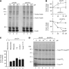
Interaction of presequence peptides with Tim50. (AF) Purified 6 × His-tagged Tim50IMS (A, B), Tim50PBD (C, D), or Tim50C (E, F) was immobilized on a Ni2+-chelator chip and the SPR response to the indicated concentrations of pALDH (A, C, E) or pALDH-s (B, D, F) was recorded. KD is presented as mean±s.e.m. (n=3 for each measurement). (G) Photocrosslinking was performed using purified 1 μM Tim50IMS, Tim50ΔPBD, Tim50C and biotinylated photopeptides pL19B and pS16B at indicated concentrations. After SDS–PAGE separation, crosslinking products were detected using streptavidin–horse radish peroxidase (SA-HRP) conjugate or by immunodecoration against Tim50. (H) HA-tagged full-length Tim50 (Tim50HA) and Tim50 fragments containing the first 365 or 361 amino acids (Tim501–365–HA, Tim501–361, and Tim501–361–HA) in pME2782 were used to replace the wild-type protein-encoding plasmid carrying URA3 in the gene deletion strain grown on 5-fluoroorotic acid-containing plates (SD-5FOA). Strains were grown on selective-Leu plates as a control.
Figure 4
Figure 4
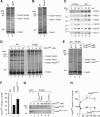
Presequence peptides affect interactions between Tim21, Tim23, and Tim50. (A) Mitochondria were pre-treated with indicated concentrations of pALDH or its inactive version pALDH-s (reaction volumes were matched by adding corresponding volumes of solvent), crosslinked by 1 mM CuSO4 for 30 min on ice and analysed by non-reducing SDS–PAGE and immunodecoration against Tim50. (B) Signals of Tim23–Tim50 (upper plot) and Tim21–Tim50 (lower plot) crosslinking-adducts (as in A) were quantified, normalized to buffer-treated controls (set as 100%), and presented as mean±s.e.m. (n=6). (C) Experiment (as in A) was performed in osmotically swollen mitochondria (mitoplasts, n=4), mitochondria with a dissipated membrane potential (–Δψ, n=4), tom22-2 (n=4) and Tim501–361–HA (n=3) mitochondria. The intensity of the Tim21–Tim50 crosslink-adducts after treatment with 50 μM pALDH was quantified and normalized to control (set to 100%). Data are presented as mean±s.e.m. (D) Purified Tim21IMS and Tim50IMS (1 μM each) were crosslinked in the presence of increasing amounts of pALDH or pALDH-s and analysed by immunoblotting against Tim21.
Figure 5
Figure 5
The IMS-domain of Tim23 facilitates interaction between Tim21IMS and Tim50. (A, B) Cu2+ crosslinking was performed in wild-type and (A) mgr2Δ or (B) Tim23 shut down mitochondria (Tim23↓), followed by immunoblotting against Tim50. (C) Indicated amounts of mitochondria from wild-type and Tim23↓ strains were analysed by SDS–PAGE and immunoblotting. (D) Mitoplasts generated from wild-type or Tim23↓ mitochondria were subjected to crosslinking with Cu2+ after incubation with the indicated concentrations of Tim21IMS. (E) Mitochondria were treated like in (D), but Tim23IMS was added where indicated prior to crosslinking. (F) Tim50–Tim21IMS crosslink intensities from independent experiments (performed as in E) after treatment with 20 μM Tim23IMS (n=9) or buffer (n=11) were quantified. Data are presented as mean±s.e.m. (G) Purified Tim21IMS was immobilized on CNBr-activated Sepharose and incubated with 20 nM Tim50IMS in the presence of the indicated Tim23IMS WT or Tim23IMS YL70AA concentrations. Bound Tim50 was eluted with 0.1 M glycin, pH 2.5 and analysed by SDS–PAGE followed by immunoblotting against Tim50. (H) Quantification of Tim50IMS signal intensities, as in (G) (presented as mean±s.e.m., n≥3).
Figure 6
Figure 6
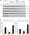
Presequences affect interactions at the TIM23 complex. (A) Co-IP was performed with Tim50 or Tim23 antisera from digitonin-solubilized mitochondria after treatment with pCox4, SynB2, or corresponding buffer. Control IP, unrelated antiserum was used as a specificity control. Eluted proteins were analysed by SDS–PAGE and immunoblotting. Total, 7%; Elution, 100%. (B) The results of seven independent IP experiments after pCox4 and SynB2 treatment utilizing Tim50 (left panel) or Tim23 (right panel) antisera (as in A) were quantified and normalized to the amount of precipitated Tim50 (for the Tim50 IP) or Tim23 (for the Tim23 IP); the SynB2 peptide control was set to 100%. Data are presented as mean±s.e.m. (n=7). The statistical significance of the changes was evaluated using a two-sided t-test. *P<0.05; **P<0.01; ***P<0.0001.
Figure 7
Figure 7
Affinities of protein–protein interactions within the presequence pathway. KD values of the interactions occurring in the mitochondrial IMS and on the matrix side of the inner mitochondrial membrane are indicated. Solid lines, affinities addressed in this study. Dashed lines, affinity data from Gevorkyan-Airapetov et al (2009); de la Cruz et al (2010); Marom et al (2011).

Similar articles

Cited by

References

    1. Abe Y, Shodai T, Muto T, Mihara K, Torii H, Nishikawa S, Endo T, Kohda D (2000) Structural basis of presequence recognition by the mitochondrial protein import receptor Tom20. Cell 100: 551–560 - PubMed
    1. Albrecht R, Rehling P, Chacinska A, Brix J, Cadamuro SA, Volkmer R, Guiard B, Pfanner N, Zeth K (2006) The Tim21 binding domain connects the preprotein translocases of both mitochondrial membranes. EMBO Rep 7: 1233–1238 - PMC - PubMed
    1. Alder NN, Sutherland J, Buhring A (2008) Quaternary structure of the mitochondrial TIM23 complex reveals dynamic association between Tim23p and other subunits. Mol Biol Cell 19: 159–170 - PMC - PubMed
    1. Allison DS, Schatz G (1986) Artificial mitochondrial presequences. Proc Natl Acad Sci USA 83: 9011–9015 - PMC - PubMed
    1. Bauer MF, Sirrenberg C, Neupert W, Brunner M (1996) Role of Tim23 as voltage sensor and presequence receptor in protein import into mitochondria. Cell 87: 33–41 - PubMed

Publication types

MeSH terms

Substances