. 2013 Apr 1;304(7):E757-69.
doi: 10.1152/ajpendo.00645.2012. Epub 2013 Feb 12.

Breast cancer resistance protein regulates apical ectoplasmic specialization dynamics stage specifically in the rat testis

Affiliations

Breast cancer resistance protein regulates apical ectoplasmic specialization dynamics stage specifically in the rat testis

Xiaojing Qian et al. Am J Physiol Endocrinol Metab. .

Abstract

Drug transporters determine the bioavailability of drugs in the testis behind the blood-testis barrier (BTB). Thus, they are crucial for male contraceptive development if these drugs (e.g., adjudin) exert their effects behind the BTB. Herein breast cancer resistance protein (Bcrp), an efflux drug transporter, was found to be expressed by both Sertoli and germ cells. Interestingly, Bcrp was not a component of the Sertoli cell BTB. Instead, it was highly expressed by peritubular myoid cells at the tunica propria and also endothelial cells of the microvessels in the interstitium at all stages of the epithelial cycle. Unexpectedly, Bcrp was found to be expressed at the Sertoli-step 18-19 spermatid interface but limited to stage VI-early VIII tubules, and an integrated component of the apical ectoplasmic specialization (apical ES). Apparently, Bcrp is being used by late-stage spermatids to safeguard their completion of spermiogenesis by preventing harmful drugs to enter these cells while they transform to spermatozoa. Also, the association of Bcrp with actin, Eps8 (epidermal growth factor receptor pathway substrate 8, an actin barbed end capping and bundling protein), and Arp3 (actin-related protein 3, a component of the Arp2/3 complex known to induce branched actin polymerization) at the apical ES suggest that Bcrp may be involved in regulating the organization of actin filament bundles at the site. Indeed, a knockdown of Bcrp by RNAi in the testis perturbed the apical ES function, disrupting spermatid polarity and adhesion. In summary, Bcrp is a regulator of the F-actin-rich apical ES in the testis.


Figures

Fig. 1.
Fig. 1.
Expression of breast cancer resistance protein (Bcrp) by Sertoli and germ cells in the rat testis. A: RT-PCR using primer pair specific to Bcrp illustrated the expression of Bcrp mRNA in the testis vs. Sertoli (SC) and germ (GC) cells. Bcrp was coamplified with S16, which served as a loading control. B: immunoblotting was performed using a specific anti-Bcrp antibody (Table 1) and lysates of adult rat testes (20 μg protein), Sertoli cells (isolated from 20-day-old rat testes and cultured for 4 days in serum-free F-12-DMEM), and germ cells (total germ cells isolated from adult rat testes and cultured for 16 h in vitro) (50 μg protein/lane for lysates of Sertoli and germ cells) to confirm findings shown in A illustrating Bcrp was expressed in the testis, but mostly restricted to germ cells and weakly expressed by Sertoli cells, with β-actin serving as a protein loading control. C: the specificity of the anti-Bcrp antibody was assessed by immunoblotting using lysates of adult rat testes (20 μg protein). Mr, relative molecular weight. D: the anti-Bcrp antibody was used for dual-labeled immunofluorescence analysis to examine the cellular localization of Bcrp (red fluorescence) vs. F-actin (green fluorescence) in the seminiferous epithelium of adult rat testes. Bcrp was localized almost exclusively to the myoid cells in the tunica propria (see “blue” arrowheads in the stage IV tubule that annotate the relative location of the tunica propria in the cross section of a seminiferous tubule), just below the blood-testis barrier (BTB) site where F-actin was highly expressed (see “white” arrowheads in the stage VI tubule that annotate the relative location of the BTB in the panel on the right), and also to the endothelial cells of the microvessels located in the interstitial space as seen in stage VII and VIII tubules (see “yellow” arrowheads in the stage VIII tubule that annotate the endothelial cells of the microvessel in the interstitium in the panel on the right). Bcrp was also expressed stage specifically at the apical ectoplasmic specialization (ES). It first appeared in stage VI tubules with relatively weak staining, and it became highly expressed in stage VII tubules, which also partially colocalized with F-actin at the apical ES. Bcrp expression diminished considerably at stage VIII tubules, and it was no longer detectable in stage IX–XI and also I–V tubules. However, Bcrp did not appear to colocalize with F-actin in the endothelial and/or smooth muscle cells of microvessels. Cell nuclei were visualized by 4′,6-diamidino-2-phenylindole (DAPI). Scale bar = 50 μm, which applies to all micrographs in this panel. E: shown is a normal adult rat testis showing the cross sections of three staged tubules at VII, VIII, and late stage VIII of the epithelial cycle using paraffin sections of testes stained by hematoxylin, which is used to illustrate the relative location of different cellular compartments being referred to in this report. The seminiferous epithelium, composed of Sertoli cells and germ cells at different stages of development (such as spermatocytes, round spermatids, and elongating/elongated spermatids), lies on the tunica propria, which is constituted by the basement membrane (composed by type IV collagen, laminin α2, heparin sulfate proteoglycan, and entactin) and type I collagen layer, to be followed by the myoid cell layer and the lymphatic endothelium (11, 41). Small blood vessels (annotated by “red” arrowheads) in the testis are restricted to the interstitium (In). Scale bar = 150 μm.
Fig. 2.
Fig. 2.
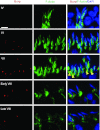
Stage-specific expression of Bcrp and its colocalization with F-actin at the apical ES during the epithelial cycle of spermatogenesis in adult rat testes. The expression of Bcrp (red fluorescence) and F-actin (green fluorescence) at the apical ES was examined by dual-labeled immunofluorescence analysis in the seminiferous epithelium of adult rat testes. Bcrp expressed stage specifically. It was first detected in stage VI tubules, and it was most prominently expressed in stage VII tubules and gradually diminished by stage VIII. Bcrp was found to be restricted almost exclusively to the concave side of step 18 (stage VI) and step 19 (stage VII–VIII) spermatid heads, colocalized with F-actin. Scale bar = 10 μm, which applies to all other micrographs in this panel.
Fig. 3.
Fig. 3.
Bcrp, an efflux drug transporter, is an integrated component of the apical ES that expressed stage specifically during the epithelial cycle. A: coimmunoprecipitation (Co-IP) was used to assess any structural interaction between Bcrp and actin, as well as a number of actin-binding and regulatory proteins. It was shown that Bcrp structurally interacted with actin, but also actin regulatory proteins epidermal growth factor receptor pathway substrate 8 (Eps8), actin-related protein 3 (Arp3), actin-related protein 2/3 complex component 2 [ARPC2; but not drebrin E (also an actin-binding protein)], palladin (an actin cross-linking and bundling protein), and several other basal ES (e.g., N-cadherin, β-catenin, afadin, JAM-C) and tight junctions (TJ) (e.g., ZO-1, occludin, JAM-A) proteins at the BTB. Data shown herein are the results of a representative experiment that was repeated three times using different rat testes and yielded similar results. (+), interaction with Bcrp was positively identified; (−), interaction with Bcrp was not identified. B and C: the association of Bcrp (red) with Eps8 (green) (B) and ARPC2 (green) (C) was further confirmed by dual-labeled immunofluorescence analysis to assess their colocalization. Colocalization between Bcrp and F-actin had been shown (see Fig. 2). Colocalization of Bcrp with Arp3 was not done because both anti-Bcrp and anti-Arp3 were mouse antibodies (see Table 1). Bcrp was found to colocalize with Eps8 and APRC2 in stage VII tubules. Similar to Bcrp, the expression of Eps8 and ARPC2 was considerably diminished in stage VIII tubules. Scale bar = 10 μm in B and C, which applies to all other micrographs in both panels.
Fig. 4.
Fig. 4.
Bcrp is not expressed at the BTB, but it is expressed by peritubular myoid cells at the tunica propria and endothelial cells of the microvessels in the interstitial space. A: dual-labeled immunofluorescence analysis of Bcrp (red) with either ZO-1 (green, a TJ adaptor protein), claudin-11 (green, a TJ integral membrane protein), or F-actin (green) in the seminiferous epithelium near the tunica propria was shown here. Bcrp was not associated with either ZO-1 or cluadin-11 in the seminiferous epithelium. In fact, Bcrp and ZO-1/claudin-11 were localized to distinctively different locations near the base of the seminiferous tubule in which Bcrp was localized to the tunica propria and associated with peritubular myoid cells (see yellow arrowheads), whereas ZO-1 and cluadin-11 were at the BTB, at the site above spermatogonia. However, Bcrp displayed partial colocalization with F-actin at the tunica propria (see “orange” arrowheads). B: Bcrp was highly expressed by endothelial cells of the microvessels in the interstitium (see white arrowheads), but it did not colocalize with F-actin in endothelial cells. Scale bar = 10 μm for A and 20 μm for B, which applies to other micrographs in the corresponding panel.
Fig. 5.
Fig. 5.
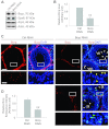
A rapid and considerable loss of Bcrp expression at the apical ES during adjudin-induced germ cell loss from the seminiferous epithelium. Rats were treated with a single dose of adjudin (50 mg/kg body wt, by gavage) to induce germ cell loss from the epithelium by disrupting apical ES function in the testis. A: immunoblot analysis to illustrate a time-dependent downregulation of Bcrp in the testis following adjudin treatment. Actin served as a protein loading control. B: this histogram summarizes results of immunoblotting shown in A, with each bar a mean ± SD of n = 3–5 rats. A time-dependent downregulation on the expression of Bcrp was noted. *P < 0.05. C: localization of Bcrp at the apical ES and its colocalization with F-actin was noted (see orange merged staining annotated by yellow arrowheads); however, a considerable downregulation of Bcrp expression at the apical ES was noted as early as 12 h after adjudin treatment. By 48 and 96 h, the expression of Bcrp at the apical ES was virtually undetectable. Scale bar = 20 μm, which applies to other micrographs in this panel. D: in contrast to the Bcrp at the apical ES, the expression of Bcrp at the tunica propria in the testis from rats following treatment of adjudin at 12, 48, and 96 h only displayed gradual downregulation, consistent with data shown in A and B. Scale bar = 20 μm, which applies to other micrographs in this panel.
Fig. 6.
Fig. 6.
Effects of Bcrp knockdown in the adult rat testis on the apical ES function. A and B: Bcrp was knocked down by RNAi with specific Bcrp siRNA duplexes (Bcrp RNAi) vs. nontargeting control duplexes (Ctrl RNAi) using seminiferous tubules isolated from adult rat testes and cultured in vitro as described in materials and methods. As shown in A, the knockdown of Bcrp in seminiferous tubules in vitro by ∼60% (B) did not lead to any off-target effects when the steady-state levels of Eps8, Arp3, and actin were assessed using 20 μg protein/lane in which actin served as a protein loading control. As shown in the histogram in B, each bar is a mean ± SD of n = 3 independent experiments using different batches of tubules isolated from adult rat testes. **P < 0.01. The experimental conditions shown in A and B were then used for studies in vivo shown in C and D in which each testis was transfected with either Bcrp specific siRNA duplexes vs. control duplexes as described in materials and methods. In short, each testis was transfected daily beginning on days 0, 1, and 2 with a total of three transfections, and rats were euthanized by CO2 asphyxiation on day 4 to assess any changes in phenotypes following the knockdown of Bcrp (C). It was noted that Bcrp was silenced by at least ∼60% (C and D) when the intensity of Bcrp signals in the seminiferous epithelium in Bcrp knockdown vs. control groups were assessed (D). C: a considerable decline in Bcrp signal at the apical ES in a stage VII tubule was noted in the Bcrp knockdown group (Bcrp RNAi) vs. the control group (Ctrl RNAi), which was also associated with a loss of spermatid adhesion (see white arrowheads, illustrating spermatids found in the tubule lumen in stage VII and early VIII tubules) and loss of spermatid polarity (see yellow arrowheads, illustrating misoriented spermatids). In stage IX tubules, elongated spermatids were found to be “trapped” in the seminiferous epithelium due to defects of spermiation because of a disruption of the apical ES function following Bcrp knockdown (see “green” arrowheads). The white boxed area in the panel on top was magnified and shown on bottom. Scale bar in the micrograph in panels on the top and bottom is 100 and 25 μm, respectively, which also apply to all other micrographs in the corresponding panel. Each bar in D is a mean ± SD of n = 60 tubules from 3 rats (20 randomly selected stage VII–VII tubules from each rat). **P < 0.01.
Fig. 7.
Fig. 7.
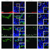
A knockdown of Bcrp in the testis impedes spermatid polarity and adhesion via changes in F-actin organization at the apical ES mediated by mislocalization of Eps8 and Arp3. A knockdown of Bcrp in the testis with Bcrp-specific siRNA duplexes (Bcrp RNAi) vs. nontargeting control duplexes (Ctrl RNAi) (see materials and methods) led to reduced Bcrp signals in the tunica propria and the apical ES in stage VII tubules when the expression of Bcrp was the highest. It was noted that a knockdown of Bcrp in these stage VII tubules by ∼60% led to loss of spermatid polarity (see yellow arrowheads). This loss of spermatid orientation was likely the result of changes in the organization of actin filament bundles at the apical ES, since F-actin was prominent at the apical ES in stage VII tubules in the Ctrl RNAi group but not in the Bcrp RNAi group. These changes in F-actin organization at the apical ES appeared to be mediated by mislocalization of: 1) Eps8 at the apical ES, which failed to maintain the actin filament bundles at the apical ES {but not at the BTB in the seminiferous epithelium, since Bcrp was not found at the BTB [see Figs. 1 and 4; see white arrowheads annotating the expression of Eps8 at the BTB in both the Bcrp RNAi and Ctrl RNAi groups, consistent with earlier reports that Eps8 is highly expressed (25) but Arp3 is downregulated (24) at the BTB in stage VII tubules]} in which Eps8 was no longer tightly localized to the concave side of spermatid heads, and also 2) Arp3 in which Arp3 was not restricted to the concave side of spermatid heads but diffused away from this site (see enlarged images in selected panels for Eps8 and Arp3 vs. Bcrp and F-actin). Boxed areas in micrographs were enlarged to illustrate changes in localization of F-actin, Eps8, and Arp3 in Bcrp RNAi vs. Ctrl RNAi groups. Cell nuclei were visualized by DAPI staining. Scale bar = 20 μm in micrograph and scale bar = 10 μm in enlarged micrograph, which apply to remaining micrographs in corresponding panels.

Similar articles

Cited by

References

    1. Ahmed S, Goh WI, Bu W. I-BAR domains, IRSp53 and filopodium formation. Semin Cell Dev Biol 21: 350–356, 2010 - PubMed
    1. Aravindan GR, Pineau C, Bardin CW, Cheng CY. Ability of trypsin in mimicking germ cell factors that affect Sertoli cell secretory function. J Cell Physiol 168: 123–133, 1996 - PubMed
    1. Bart J, Hollema H, Groen HJ, de Vries EG, Hendrikse NH, Sleijfer DT, Wegman TD, Vaalburg W, van der Graaf WT. The distribution of drug-efflux pumps, P-gp, BCRP, MRP1 and MRP2, in the normal blood-testis barrier and in primary testicular tumours. Eur J Cancer 40: 2064–2070, 2004 - PubMed
    1. Campbell M, Humphries P. The blood-retina barrier: tight junctions and barrier modulation. Adv Exp Med Biol 763: 70–84, 2012 - PubMed
    1. Chen YM, Lee NPY, Mruk DD, Lee WM, Cheng CY. Fer kinase/Fer T and adherens junction dynamics in the testis: an in vitro and in vivo study. Biol Reprod 69: 656–672, 2003 - PubMed

Publication types

MeSH terms

LinkOut - more resources