. 2013 Mar;140(5):1100-10.
doi: 10.1242/dev.088989.

The tiptop/teashirt genes regulate cell differentiation and renal physiology in Drosophila

Affiliations

The tiptop/teashirt genes regulate cell differentiation and renal physiology in Drosophila

Barry Denholm et al. Development. 2013 Mar.

Abstract

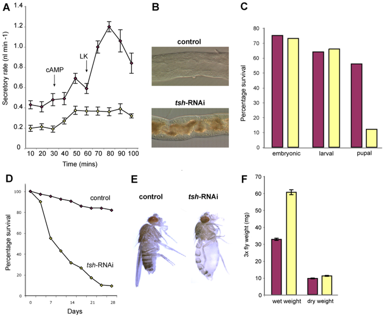
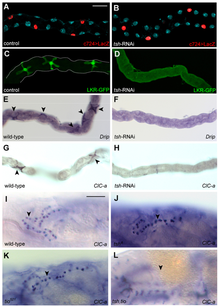
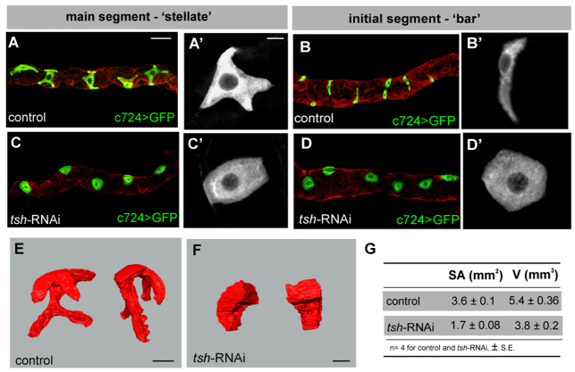
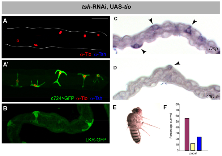
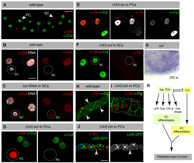
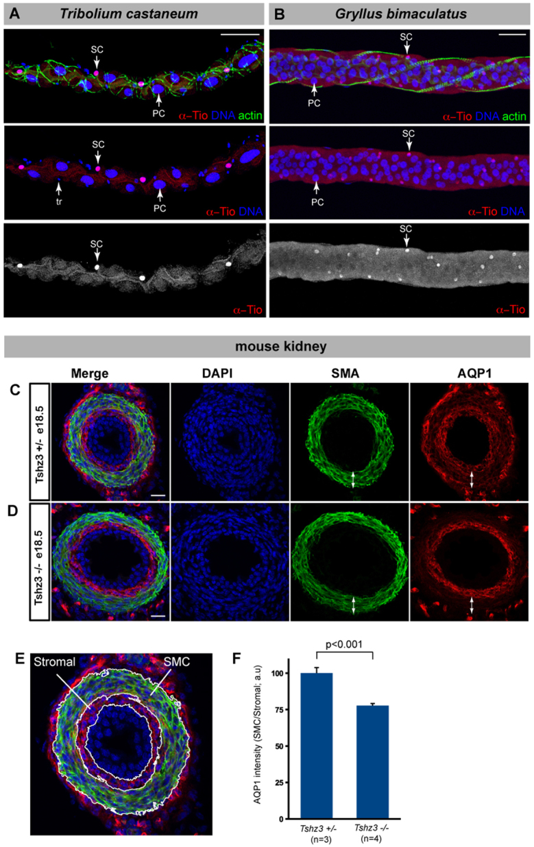
The physiological activities of organs are underpinned by an interplay between the distinct cell types they contain. However, little is known about the genetic control of patterned cell differentiation during organ development. We show that the conserved Teashirt transcription factors are decisive for the differentiation of a subset of secretory cells, stellate cells, in Drosophila melanogaster renal tubules. Teashirt controls the expression of the water channel Drip, the chloride conductance channel CLC-a and the Leukokinin receptor (LKR), all of which characterise differentiated stellate cells and are required for primary urine production and responsiveness to diuretic stimuli. Teashirt also controls a dramatic transformation in cell morphology, from cuboidal to the eponymous stellate shape, during metamorphosis. teashirt interacts with cut, which encodes a transcription factor that underlies the differentiation of the primary, principal secretory cells, establishing a reciprocal negative-feedback loop that ensures the full differentiation of both cell types. Loss of teashirt leads to ineffective urine production, failure of homeostasis and premature lethality. Stellate cell-specific expression of the teashirt paralogue tiptop, which is not normally expressed in larval or adult stellate cells, almost completely rescues teashirt loss of expression from stellate cells. We demonstrate conservation in the expression of the family of tiptop/teashirt genes in lower insects and establish conservation in the targets of Teashirt transcription factors in mouse embryonic kidney.


Figures

Fig. 1.
Fig. 1.
Drosophila Malpighian tubules. (A) The four MpTs consist of initial, transitional and main segments, and a ureter. Stellate cells (SCs, green) are interspersed with principal cells (PCs, yellow) in the initial and transitional segments (where they are bar shaped), and throughout the secretory region of the main segment (where they are stellate). (B) The major physiological activities carried out by PCs (yellow) and SCs (green). Ion transport is driven by a H+-transporting vacuolar-ATPase (V-ATPase) on the luminal membrane of PCs, which, coupled with a cation/H+ antiporter, transports potassium ions into the lumen. Chloride ions move down an electrochemical gradient through chloride channels in SCs. Water (blue arrows) follows by osmosis through water channels in SCs and paracellular routes. Capability peptides 1/2 (Capa) and diuretic hormone 31/44 (DH) stimulate urine production through cGMP and cAMP pathways in PCs (Cabrero et al., 2002; Coast et al., 2001; Johnson et al., 2005; Kean et al., 2002). BM, basement membrane; LK, leucokinin; LKR, leucokinin receptor; SJ, septate junction.
Fig. 2.
Fig. 2.
Tsh and Tio expression in Drosophila tubules. (A-F) Tsh (red, A-C) and Tio (black, D-F) expression in embryonic MpTs. Tsh is first detected in SCs at stage 13 (B, arrow) and maintained throughout embryogenesis. Tio is first detected in PCs from stage 11 (D, arrows) and in SCs from stage 13 (E). By stage 16, Tio expression levels in SCs are high but diminished in PCs (F). (G,H) Low- (G) and high- (H) magnification views of stage 16 embryo showing Tsh (red) and Tio (green, overlap appears yellow) co-expression in SCs (arrows). (I-L) Tsh (red; I,J) and Tio (red; K,L) expression in larval (third instar; I,K) and adult (J,L) MpTs. Tsh but not Tio is expressed in larval and adult SCs. PCs are marked with anti-Cut (green, A-C) and SCs with c724>CD8GFP (green, I-L). Tubule outline is marked with broken lines. Scale bars: 50 μm in A-F; 50 μm in G,H; 30 μm in I-L.
Fig. 3.
Fig. 3.
Disrupted organ function and animal physiology in tsh knockdown flies. (A) Secretory rates (nl min–1) in control (purple) and tsh knockdown (yellow) MpTs. Cyclic adenosine monophosphate (cAMP) and Leucokinin (LK) were added at ∼30 and 60 minutes, respectively (arrows). Basal secretion rates are lower, and the response to LK is abolished in tsh knockdown tubules. (B) Uric acid crystals accumulate in the MpT lumen in the tsh knockdown. (C) Survival rates of control (purple, n=600) and tsh knockdown (yellow, n=900, nine replicates) animals from embryonic to pupal stages. The main lethal phase in tsh knockdown occurs during pupation. (D) Survival rates for control (purple, n=105) and tsh knockdown (yellow, n=140) adults. (E) Control (left) and tsh knockdown (right) 1-week-old adults. tsh knockdown adults have grossly distended abdomens. (F) Wet and dry weight measurements (mg; three flies/measurement) for adults: control (purple, n=13); tsh knockdown (yellow, n=26). Data are for females (equivalent results for males not shown).
Fig. 4.
Fig. 4.
tsh regulates terminal differentiation gene expression. (A,B) SC number and spacing is normal in control (A) and tsh knockdown (B) adult MpTs. SCs marked with c724>UAS-nlacZ (red), tubules counterstained for DNA (blue). (C-H) tsh regulates SC gene expression in MpTs. Adult MpTs from control or wild-type (C,E,G), or tsh knockdown (D,F,H) animals. (C,D) LKR-GFP (LKR protein trap), (E,F) Drip and (G,H) ClC-a in situ hybridisation. LKR, Drip and ClC-a expression is completely abolished in tsh knockdown SCs. Arrowheads in E,G. (I-L) tsh and tio regulate ClC-a expression redundantly in embryonic SCs. ClC-a expression (in situ hybridisation) in stage 17 embryos in wild type (I), tsh mutant (J), tio mutant (K) and tsh tio double mutant (L). ClC-a expression in embryonic SCs is unaffected in either tsh or tio mutants (arrowheads, I-K), but completely abolished in tsh tio double mutants (arrowhead indicates tubule position, L). Scale bars: 30 μm in A-H; 50 μm in I-L.
Fig. 5.
Fig. 5.
tsh is required for SC morphology. (A-D′) Adult MpTs, SCs marked with c724>UAS-CD8GFP (green in A-D, white in A′-D′); tubules counterstained for actin (phalloidin, red). (A,B) Control tubules showing ‘stellate’-shaped SCs in main segment (A,A′) and ‘bar’-shaped cells in initial segment (B,B′). (C,D) tsh knockdown tubules have uncharacteristically round SCs in main (C,C′) and initial (D,D′) segments. (E,F) Three-dimensional reconstruction of control (E) and tsh knockdown (F) SCs shown from different angles. (G) Surface area and volume measurements for control (n=4) and tsh knockdown (n=4) SCs. (The reduction in volume accounts for only a 20% reduction in surface area in tsh knockdown SCs.) Scale bars: 30 μm in A-D; 10 μm in A′-D′,E,F.
Fig. 6.
Fig. 6.
tio is functionally equivalent to tsh. (A) Tio (red) and Tsh (blue) expression in SCs in c724>tsh-RNAi, UAS-tio, UAS-GFP adult MpT. Tio is expressed at high levels, whereas Tsh is undetectable. (A′) SCs morphology (c724>GFP, green) is rescued by Tio (red) in the absence of Tsh (blue). (B-F) Tio rescues SC gene expression, fluid homeostasis and lethality in the absence of Tsh. (B-D) Adult MpTs from c724>tsh-RNAi, UAS-tio animals LKR (B, LKR protein trap, green), and Drip (C) and ClC-a (D) in situ hybridisation. Arrowheads indicate SCs. (E) Adult c724>tsh-RNAi, UAS-tio do not exhibit fluid a homeostasis phenotype. (F) Survival rates of control (purple), knockdown (yellow) and c724>tsh-RNAi, UAS-tio (blue) expressed as percentage of adult hatching from 600, 900 and 324 embryos (four replicates carried out for tio rescue). Tio partly rescues the lethality associated with tsh knockdown. Scale bars: 30 μm in A-D.
Fig. 7.
Fig. 7.
The tsh gene network in SCs. (A) Wild-type adult tubule showing mutually exclusive expression of Cut (green) in PCs (large nuclei) and Tsh (red) in SCs (small nuclei). (B,C) Adult tubules stained for Cut (red) and DNA (white). In wild type (B), Cut is expressed in PCs (large nuclei) but not in SCs (small nucleus, circled). In tsh knockdown (C), Cut expression is ectopically induced in SCs (small nucleus, circled). (D) Adult tubule with induced ectopic expression of tsh in PCs (CtB>UAS-tsh results in mosaic tsh expression in PCs) stained for Cut (green) and Tsh (red). Ectopic Tsh in PCs (circled) leads to repression of Cut. (E) Adult tubule with ectopic expression of tio in PCs (CtB>UAS-tio results in mosaic and variable levels of tio expression in PCs) stained for Cut (green, white in individual channels) and Tio (red, white in individual channels). Ectopic tio expression in PCs leads to repression of Cut in a dose-dependent manner; high (+++), medium (++) and low (+) levels of Tio are indicated. (F) Adult tubule with ectopic expression of cut in SCs (c724>UAS-cut). Ectopic Cut expression leads to repression of Tsh in SCs (circled). (G) ClC-a in situ hybridisation in a cut mutant embryo. ClC-a is not ectopically expressed in PCs in the absence of Cut. The blister-shaped MpTs are outlined. (H,I) Adult MpTs stained for Disc-large (Dlg, green) to reveal cell shape and Tsh (red). Tsh-expressing PCs are transformed to a bar-shaped morphology (I) compared with control PCs (arrowheads, H). (J) Adult tubule with ectopic expression of tsh in PCs (CtB>UAS-tsh results in mosaic tsh expression in PCs) stained for Cut (blue), Tsh (red) and LKR-GFP (green, white in single channel image). LKR expression is not induced in Tsh-expressing PCs. (K) Schematic drawing of the gene network controlling PC and SC differentiation. Scale bars: 30 μm in A,H,I; 10 μm in B-F; 50 μm in G,J.
Fig. 8.
Fig. 8.
Conservation of Tsh function. (A,B) MpTs from adult beetle (Tribolium castaneum) and cricket (Gryllus bimaculatus) stained with an antibody against D.m. Tio (red, white in single channel), DNA (blue) and actin (phalloidin, green). Tio marks a subset of MpTs cells with small nuclei interspersed with cells with larger nuclei. [In A, cells with the smallest nuclei (tr) correspond to tracheal cells.] (C-E) Cross-section through E18.5 mouse ureter stained for DAPI (blue), smooth muscle actin (SMA, green) and aquaporin 1 (AQP1, red), in sibling control (C) or Tshz3 homozygote (D). Double-headed arrows indicate smooth muscle layer. AQP1 expression in the smooth muscle layer is reduced in Tshz3–/– ureters. (E) Stromal and smooth muscle layer (SMC) are indicated in an enlarged image from C. (F) Quantification of AQP1 expression in the SMC by fluorescence intensity (expression in SMC was normalised relative to expression in the stromal layer). Both the wild-type and mutant ureters were sectioned distal to the ureteropelvic junction because of the severe hydroureter phenotype in the mutant ureter (Caubit et al., 2008). Scale bars: 50 μm in A,B; 25 μm in C,D.

Similar articles

Cited by

References

    1. Al-Anzi B., Armand E., Nagamei P., Olszewski M., Sapin V., Waters C., Zinn K., Wyman R. J., Benzer S. (2010). The leucokinin pathway and its neurons regulate meal size in Drosophila. Curr. Biol. 20, 969–978 - PMC - PubMed
    1. Alexandre E., Graba Y., Fasano L., Gallet A., Perrin L., De Zulueta P., Pradel J., Kerridge S., Jacq B. (1996). The Drosophila teashirt homeotic protein is a DNA-binding protein and modulo, a HOM-C regulated modifier of variegation, is a likely candidate for being a direct target gene. Mech. Dev. 59, 191–204 - PubMed
    1. Andrew D. J., Horner M. A., Petitt M. G., Smolik S. M., Scott M. P. (1994). Setting limits on homeotic gene function: restraint of Sex combs reduced activity by teashirt and other homeotic genes. EMBO J. 13, 1132–1144 - PMC - PubMed
    1. Azpiazu N., Morata G. (2000). Function and regulation of homothorax in the wing imaginal disc of Drosophila. Development 127, 2685–2693 - PubMed
    1. Berridge M. J., Oschman J. L. (1969). A structural basis for fluid secretion by malpighian tubules. Tissue Cell 1, 247–272 - PubMed

Publication types

MeSH terms