. 2013 Mar 11;54(3):1816-28.
doi: 10.1167/iovs.12-10741.

Delta-opioid agonist SNC-121 protects retinal ganglion cell function in a chronic ocular hypertensive rat model

Affiliations

Delta-opioid agonist SNC-121 protects retinal ganglion cell function in a chronic ocular hypertensive rat model

Yasir Abdul et al. Invest Ophthalmol Vis Sci. .

Abstract

Purpose: This study examined if the delta-opioid (δ-opioid) receptor agonist, SNC-121, can improve retinal function and retinal ganglion cell (RGC) survival during glaucomatous injury in a chronic ocular hypertensive rat model.

Methods: IOP was raised in brown Norway rats by injecting hypertonic saline into the limbal venous system. Rats were treated with 1 mg/kg SNC-121 (intraperitoneally [IP]) once daily for 7 days. Pattern-electroretinograms (PERGs) were obtained in response to contrast reversal of patterned visual stimuli. RGCs were visualized by fluorogold retrograde labeling. Expression of TNF-α and p38 mitogen-activated protein (MAP) kinase was measured by immunohistochemistry and Western blotting.

Results: PERG amplitudes in ocular hypertensive eyes were significantly reduced (14.3 ± 0.60 μvolts) when compared with healthy eyes (18.0 ± 0.62 μvolts). PERG loss in hypertensive eyes was inhibited by SNC-121 treatment (17.20 ± 0.1.3 μvolts; P < 0.05). There was a 29% loss of RGCs in the ocular hypertensive eye, which was inhibited in the presence of SNC-121. TNF-α production and activation of p38 MAP kinase in retinal sections and optic nerve samples were upregulated in ocular hypertensive eyes and inhibited in the presence of SNC-121. Furthermore, TNF-α induced increase in p38 MAP kinase activation in astrocytes was inhibited in the presence of SNC-121.

Conclusions: These data provide evidence that activation of δ-opioid receptors inhibited the loss of PERG amplitudes and rate of RGC loss during glaucomatous injury. Mechanistic data provided clues that TNF-α is mainly produced from glial cells and activates p38 MAP kinase, which was significantly inhibited by SNC-121 treatment. Overall, data indicate that enhancement of δ-opioidergic activity in the eye may provide retina neuroprotection against glaucoma.


Conflict of interest statement

Disclosure: Y. Abdul, None; N. Akhter, None; S. Husain, None

Figures

Figure 1
Figure 1
IOP measurements of healthy eyes, and eyes from a chronic glaucoma model with and without SNC-121 (1 mg/kg) treatment for 7 days, once daily. Rats were divided into two groups: ocular hypertensive group (n = 8) and SNC-121–treated ocular hypertensive group (n = 8). IOP was elevated in one eye of brown Norway rats by injecting approximately 50 μL of 2.0 M hypertonic saline, while the contralateral eye served as the control. Rats were maintained for up to 6 weeks post surgery. A total of 16 rats were used in this experiment. Data are mean ± SE. *P < 0.05; n = 8 for each group.
Figure 2
Figure 2
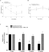
(A) PERG recording in normal and ocular hypertensive rat eyes. Each waveform is a mean of 300 individual waveforms taken at an interval of 1 second for each data point. (B) Example of PERG recorded in SNC-121–treated (1 mg/kg) healthy and ocular hypertensive rat eyes. Each waveform is also a mean of 300 individual waveforms. (C) Changes observed in PERG of untreated and SNC-121–treated ocular hypertensive rat eyes. In these experiments, brown Norway rats were treated with 1 mg/kg SNC-121 (IP) immediately after hypertonic saline injections, and subsequently once daily for 7 days. PERG data shown in this figure were collected at weeks 2, 4, and 6, post saline injection. Prior to the surgery, PERG values in both hypertensive and SNC-121–treated hypertensive eyes (e.g., ranging from 16–20 μvolts) were considered as 100% at day 0. The changes in PERG of hypertensive eyes or SNC-121–treated eyes at 2, 4, and 6 weeks were calculated using their respective values at day 0. A total of 15 rats (e.g., ocular hypertensive group, n = 8 and SNC-121–treated ocular hypertensive group, n = 7) were used in this experiment. IOP data for these rats is shown in Figure 1. Data are mean ± SE; *P < 0.05; n = 7 to 8.
Figure 3
Figure 3
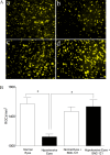
(A) Fluorescence micrographs of flat-mounted retinas depicting Fluorogold-labeled RGCs in normal (a), ocular hypertensive (b), SNC-121–treated normal (c), and SNC-121–treated ocular hypertensive (d), eyes. Briefly, 3 μL of a 5% solution of fluorogold was injected into the superior colliculus of anesthetized animals. Seven days post injection, animals were euthanized and retinas were prepared as flat-mounts, vitreous side facing up. Fluorescent RGCs were visualized under Zeiss microscopy. Bar: 20 μm. (B) Quantification of RGCs. Rats were divided into two groups: ocular hypertensive group (n = 6) and SNC-121–treated ocular hypertensive group (n = 6). A total of 12 rats were used in this experiment. IOP and PERG data for these rats are shown in Figures 1 and 2, respectively. RGCs were counted in eight microscopic fields of identical size (150 μm2 area) for each retina using ImageJ software. *P < 0.05; n = 6 for each group.
Figure 4
Figure 4
Eyes of brown Norway rats were enucleated 7 days post hypertonic saline injections. Contralateral eyes were used as the normal control. Cryosections were immunostained by anti–TNF-α and anti-GFAP antibodies as indicated horizontally. Ocular treatments are indicated vertically. Green color indicates staining for TNF-α, red for GFAP, and blue nuclei for DAPI. Far right panels represent double-labeling of TNF-α and GFAP. There was no positive staining when primary antibodies were omitted (not shown). Fluorescence microscopy; bar is 20 μm. Data shown in this figure are a representation of at least four independent experiments. A total of 10 animals were used in this experiment. Comparable staining for TNF-α and GFAP was seen in at least four animals in each treatment group.
Figure 5
Figure 5
Eyes of brown Norway rats were enucleated 7 days post hypertonic saline injection. Contralateral eyes were used as the healthy control. Cryosections were immunostained by anti–TNF-α antibodies and anti-CRALBP, as indicated horizontally. Ocular treatments are indicated vertically. Green color indicates staining for TNF-α, red for CRALBP, and blue nuclei for DAPI. Far right panels represent double-labeling of TNF-α and CRALBP. There was no positive staining when primary antibodies were omitted (not shown). Fluorescence microscopy; bar is 20 μm. Data shown in this figure are representative of at least four independent experiments. A total of 10 animals were used in this experiment. Comparable staining for TNF-α and CRALBP was seen in at least four animals in each treatment group.
Figure 6
Figure 6
TNF-α production in response to glaucomatous injury and its suppression by SNC-121 treatment in the optic nerve extracts. Optic nerves of untreated and SNC-121–treated healthy and hypertensive eyes were collected at 3, 7, or 42 days post injury and analyzed for TNF-α expression by Western blotting using anti–TNF-α antibodies. Rats were divided into two groups: ocular hypertensive group (n = 4) and SNC-121–treated ocular hypertensive group (n = 4). Rats were treated with SNC-121 (1 mg/kg) once daily for 7 days. IOP was elevated in one eye of brown Norway rats by injecting approximately 50 μL of 2.0 M hypertonic saline into limbal veins, while the contralateral eye served as the control. Band intensities of Western blots were quantitated by densitometry. A total of 24 animals were used in this experiment. Data are expressed as mean ± SE. *P < 0.05; n = 4 for each group.
Figure 7
Figure 7
The ONH of brown Norway rats was removed 42 days post hypertonic saline injection. Contralateral optic nerve was used as the normal control. Cryosections were immunostained by anti–TNF-α antibodies as indicated horizontally. Ocular treatments are indicated vertically. Green color indicates staining for TNF-α and blue nuclei for DAPI. There was no positive staining when primary antibodies were omitted (not shown). Data shown in this figure are a representation of at least four independent experiments. A total of eight animals were used in this experiment. Comparable staining for TNF-α was seen in at least four animals in each treatment group.
Figure 8
Figure 8
Eyes of brown Norway rats were enucleated 7 days post hypertonic saline injection. Contralateral eyes were used as the healthy control. Cryosections were immunostained by anti–phospho-p38 antibodies and anti-GFAP as indicated horizontally. Ocular treatments are indicated vertically. Green color indicates staining for phospho-p38, red for GFAP, and blue nuclei for DAPI. Far right panels show double labeling of phospho-p38 and GFAP. There was no positive staining when primary antibodies were omitted (not shown). Fluorescence microscopy; bar is 20 μm. A total of 10 animals were used in this experiment. Data shown in this figure are a representation of at least five independent experiments.
Figure 9
Figure 9
Activation of p38 MAP kinase in response to glaucomatous injury and its suppression by SNC-121 treatment in the optic nerve extracts. Optic nerves of untreated and SNC-121 treated healthy and ocular hypertensive eyes were collected at 3, 7, or 42 days post injury. Changes in phosphorylation of p38 MAP kinase were analyzed by Western blotting using selective anti–phospho-p38 MAP kinase antibodies. Rats were divided into two groups: ocular hypertensive group (n = 6) and SNC-121–treated ocular hypertensive group (n = 6). IOP was elevated in one eye of brown Norway rats by injecting approximately 50 μL of 2.0 M hypertonic saline, while the contralateral eye served as the control. A total of 36 animals were used in this experiment. Band intensities of Western blots were quantitated by densitometry. Data are expressed as mean ± SE. *P < 0.05; n = 6 for each group at each time point.
Figure 10
Figure 10
TNF-α–induced activation of p38 MAP kinase in ONH astrocytes. ONH astrocytes were starved in serum-free medium for overnight. Cells were then pretreated with SNC-121 (1 μmol/L) for 15 minutes followed by TNF-α (25 ng/mL) treatment for 6 hours. Cell lysate (15 μg protein) was analyzed by Western blotting using selective anti–phospho-p38 MAP kinase antibodies followed by incubation with appropriate secondary antibodies (HRP-conjugated; dilution 1:3000). The signal was captured using enhanced chemiluminescent reagent and the Biorad Versadoc imaging system. Data shown is a representative of four independent experiments. Data are expressed as mean ± SE. *P < 0.05; n = 4.
Figure 11
Figure 11
Determination of δ-opioid receptors in healthy and ocular hypertensive rat retinas at 3 (A) and 7 days (B) post injury. Retinal extract (15–20 μg proteins) were analyzed by Western blotting using selective anti–δ antibodies followed by incubation with appropriate secondary antibodies (HRP-conjugated; dilution 1:3000). The signal was captured using enhanced chemiluminescent reagent and the Biorad Versadoc imaging system. A total of 14 rats were used in this experiment. Data shown are representative of seven independent experiments. Data are expressed as mean ± SE. *P < 0.05; n = 7 for each time point.

References

    1. Quigley HA, Green WR. The histology of human glaucoma cupping and optic nerve damage: clinicopathologic correlation in 21 eyes. Ophthalmology. 1979; 86: 1803–1830 - PubMed
    1. Boland MV, Quigley HA. Risk factors and open-angle glaucoma: classification and application. J Glaucoma. 2007; 16: 406–418 - PubMed
    1. Liu Q, Ju WK, Crowston JG, et al. Oxidative stress is an early event in hydrostatic pressure induced retinal ganglion cell damage. Invest Ophthalmol Vis Sci. 2007; 48: 4580–4589 - PubMed
    1. Ju WK, Kim KY, Angert M, et al. Memantine blocks mitochondrial OPA1 and cytochrome c release and subsequent apoptotic cell death in glaucomatous retina. Invest Ophthalmol Vis Sci. 2009; 50: 707–716 - PMC - PubMed
    1. Nucci C, Tartaglione R, Rombola L, Morrone LA, Fazzi E, Bagetta G. Neurochemical evidence to implicate elevated glutamate in the mechanisms of high intraocular pressure (IOP)-induced retinal ganglion cell death in rat. Neurotoxicology. 2005; 26: 935–941 - PubMed

Publication types

MeSH terms

LinkOut - more resources